. 2007 Oct;6(10):1824-40.
doi: 10.1128/EC.00188-07. Epub 2007 Aug 22.

Role of actin cytoskeletal dynamics in activation of the cyclic AMP pathway and HWP1 gene expression in Candida albicans

Affiliations

Role of actin cytoskeletal dynamics in activation of the cyclic AMP pathway and HWP1 gene expression in Candida albicans

Michael J Wolyniak et al. Eukaryot Cell. 2007 Oct.

Abstract

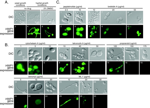
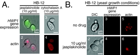
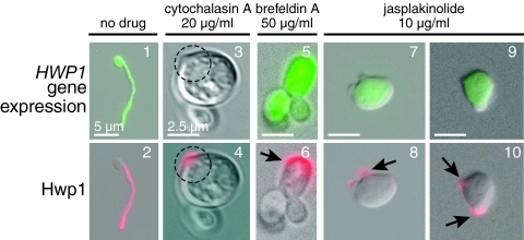
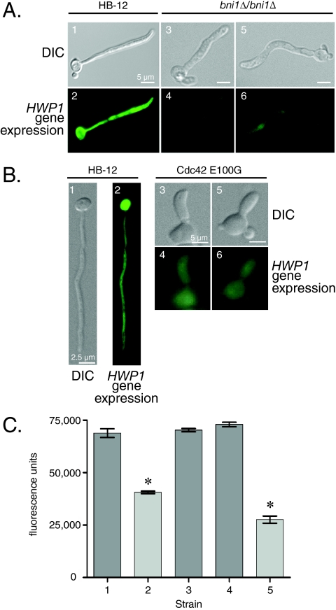
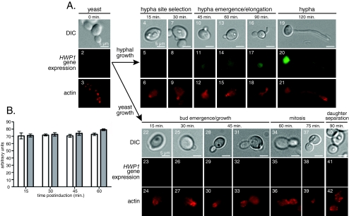
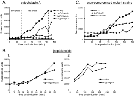
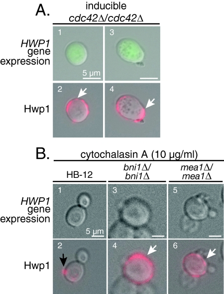
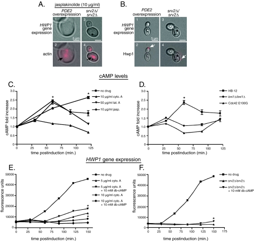
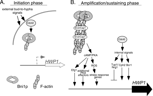
Changes in gene expression during reversible bud-hypha transitions of the opportunistic fungal pathogen Candida albicans permit adaptation to environmental conditions that are critical for proliferation in host tissues. Our previous work has shown that the hypha-specific adhesin gene HWP1 is up-regulated by the cyclic AMP (cAMP) signaling pathway. However, little is known about the potential influences of determinants of cell morphology on HWP1 gene expression. We found that blocking hypha formation with cytochalasin A, which destabilizes actin filaments, and with latrunculin A, which sequesters actin monomers, led to a loss of HWP1 gene expression. In contrast, high levels of HWP1 gene expression were observed when the F-actin stabilizer jasplakinolide was used to block hypha formation, suggesting that HWP1 expression could be regulated by actin structures. Mutants defective in formin-mediated nucleation of F-actin were reduced in HWP1 gene expression, providing genetic support for the importance of actin structures. Kinetic experiments with wild-type and actin-deficient cells revealed two distinct phases of HWP1 gene expression, with a slow, actin-independent phase preceding a fast, actin-dependent phase. Low levels of HWP1 gene expression that appeared to be independent of stabilized actin and cAMP signaling were detected using indirect immunofluorescence. A connection between actin structures and the cAMP signaling pathway was shown using hyper- and hypomorphic cAMP mutants, providing a possible mechanism for up-regulation of HWP1 gene expression by stabilized actin. The results reveal a new role for F-actin as a regulatory agent of hypha-specific gene expression at the bud-hypha transition.


Figures

FIG. 1.
FIG. 1.
HWP1 gene expression in the presence of drug treatments to disrupt hyphal morphology. (A) C. albicans strain HB-12, containing GFP under the control of the HWP1 promoter, was incubated in conditions supporting yeast (1 and 2) or hyphal (3 to 6) growth. The addition of 2% dimethyl sulfoxide (5 and 6) had no effect on hypha formation or GFP fluorescence. (B) Drug treatments that disrupted hyphal growth with loss of HWP1 gene expression: cytochalasin A (1 to 6), latrunculin A (7 to 12), propranolol (13 to 18), benomyl (19 to 24), and ML-7 (25 to 30). (C) Drug treatments that preserved HWP1 gene expression despite the loss of hypha formation included jasplakinolide (1 to 4) and brefeldin A (5 to 10). Note that fluorescence reflecting HWP1 gene expression upon loss of hyphal morphology is intense with jasplakinolide at 10 μg/ml and is barely detectable with cytochalasin A at 10 μg/ml in the dark-field image (B, 6). DIC microscopy (odd-numbered panels) and epifluorescence microscopy (even-numbered panels) were used to detect morphology and HWP1 gene expression, respectively. Drugs were used at the indicated concentrations.
FIG. 2.
FIG. 2.
(A) Correlation between HWP1 gene expression and the presence of aggregated actin structures in strain HB-12 treated with 10 μg/ml cytochalasin A or jasplakinolide under hypha-inducing conditions. F-actin was detected by staining with rhodamine-conjugated phalloidin. F-actin was aggregated following treatment with 10 μg/ml jasplakinolide (2, arrow) but not with 10 μg/ml cytochalasin A (4). GFP fluorescence reflecting HWP1 gene expression was observed in the presence of jasplakinolide (1) but not cytochalasin A (3). (B) HWP1 gene expression was absent in cells grown under yeast conditions in the presence or absence of 10 μg/ml jasplakinolide.
FIG. 3.
FIG. 3.
Hwp1 localization in the presence of disrupted hyphal morphology. Hwp1 was detected on hyphal cell surfaces of strain HB-12 in the absence (1 and 2) or presence of cytochalasin A (3 and 4), brefeldin A (5 and 6), or jasplakinolide (7 through 10) by indirect immunofluorescence using anti-Hwp1 primary antibody and Texas Red-conjugated goat anti-rabbit secondary antibody. Drugs were used at the indicated concentrations. Fluorescence produced by the GFP reporter reflected HWP1 gene expression. Note the putative site of germ tube formation (3, dashed circle) and the aggregation of Hwp1 in this region (4, dashed circle) in the cytochalasin A-treated cell, in which HWP1 gene expression, as assessed by measuring the fluorescence of GFP, was not detectable. Hwp1 was confined to localized regions of the cell surface of brefeldin A (6, arrow)- and jasplakinolide (8 and 10, arrows)-treated cells. Images 3 to 10 are shown at the same scale as image 3.
FIG. 4.
FIG. 4.
HWP1 gene expression in mutants defective for actin nucleation. C. albicans strains CKFY254 (pfy1Δ/pfy1Δ), CKFY 354 (bnr1Δ/bnr1Δ), MWY1 (bni1Δ/bni1Δ), and AV03 (Cdc42 E100G) were transformed with p1902GFP3NAT1 to monitor HWP1 gene expression in comparison to strain HB-12 (A and B). Microscopic analysis of hyphae by DIC (1, 3, and 5) and epifluorescence (2, 4, and 6) microscopy was performed to detect HWP1 gene expression in strains HB-12 and MWY1 (A) and AV03 (B). Note the decreased fluorescence of the bni1Δ/bni1Δ (A, 4 and 6) and Cdc42 E100G (B, 4 and 6) strains compared to that of HB-12 (2), an effect that was not observed in bnr1Δ/bnr1Δ and pfy1Δ/pfy1Δ cells (not shown). (C) Fluorometry to quantitate HWP1 gene expression in the bni1Δ/bni1Δ and Cdc42 E100G strains (strains 2 and 5; light bars) and in the HB-12, bnr1Δ/bnr1Δ, and pfy1Δ/pfy1Δ strains (strains 1, 3, and 4; dark bars). *, P < 0.001 (bni1Δ/bni1Δ strain versus HB-12 and Cdc42 E100G strain versus HB-12).
FIG. 5.
FIG. 5.
(A) Correlation between hypha formation, actin polarization, and HWP1 gene expression. Cells were grown in hypha (4 to 21)- or yeast (22 to 42)-inducing conditions and examined for morphology (top rows), HWP1 gene expression (middle rows), and rhodamine-phalloidin stained F-actin (bottom rows). A different individual cell is shown at each time point. (B) Quantitation of F-actin prior to germ tube and bud emergence. The amount of aggregated F-actin is shown for hyphal (gray bars) and yeast (white bars) cells.
FIG. 6.
FIG. 6.
Role of F-actin in the kinetics of HWP1 gene expression. (A) Cytochalasin A (cyto. A)-treated HB-12. (B) Jasplakinolide-treated (10 μg/ml) HB-12 cells in the slow (1) or fast (2) phase of HWP1 gene activation. (C) Strains MWY1 (bni1Δ/bni1Δ) and AV03 (Cdc42 E100G). Each graph in panels A to C is a representative example of experiments that were repeated three times.
FIG. 7.
FIG. 7.
Presence of HWP1 gene expression in cells made defective for cell surface polarization using chemical and genetic methods. Interference with cell surface polarization was accomplished using SMC7A grown under conditions leading to the loss of CDC42 (A) or using strains MWY1 (bni1Δ/bni1Δ) and MWY2 (mea1Δ/mea1Δ) that had been treated with cytochalasin A (B). Strain HB-12 served as a wild-type control. Strain HB-12 showed a punctate distribution of Hwp1 (B, 2, black arrow), in contrast to cells in which polarization was inhibited (B, 4 and 6, white arrows).
FIG. 8.
FIG. 8.
Role of cAMP in actin-mediated HWP1 gene expression. (A) HWP1 gene expression and F-actin staining in EPDE2-3 (PDE2 overexpression) (1 and 2) and MWY3 (srv2Δ/srv2Δ) (3 and 4) strains exposed to 10 μg/ml jasplakinolide under hypha-inducing conditions. (B) Localization of Hwp1 to a distinct region on cell surfaces of the srv2Δ/srv2Δ and PDE2 overexpression strains (2 and 4, arrows). HWP1 gene expression was insufficient to be detectable by measuring the fluorescence of the GFP reporter in both genetic backgrounds (1 and 3). (C) Intracellular cAMP levels of cells treated with actin-influencing agents. *, P < 0.001 (no drug versus 10 μg/ml cytochalasin A [cyto. A] at 60 min and no drug versus 10 μg/ml cytochalasin A, latrunculin A, and jasplakinolide at 120 min). (D) Intracellular cAMP levels of actin-compromised mutant cells. *, P < 0.001 (no drug versus both mutant strains at 60 min). (E) HWP1 gene expression in the presence of 5 or 10 μg/ml cytochalasin and the presence or absence of 10 mM dibutyryl (db)-cAMP. *, P < 0.001 (5 μg/ml and 10 μg/ml cytochalasin A plus db-cAMP versus no dibutyryl-cAMP, at 150 min). (F) HWP1 gene expression of strain MWY3 (srv/srv2Δ) in the presence (inverted triangles) or absence (triangles) of 10 mM dibutyryl-cAMP. *, P < 0.01 (with dibutyryl-cAMP versus no dibutyryl-cAMP at 150 min).
FIG. 9.
FIG. 9.
Model for biphasic expression and up-regulation of HWP1 at the bud-hypha transition. (A) In the initial-expression phase, environmental signals activate undetermined signal transduction pathways, allowing for small-scale HWP1 gene expression as shown by the gray arrow. Stabilized actin is not required for this gene expression. This initial HWP1 expression occurs simultaneously with polarization events resulting from Cdc42 interactions with Bni1 in conjunction with other proteins, such as Mea1p, that lead to localized Hwp1 deposition on the cell wall surface. While Cdc42 interactions with Bni1 may be promoting formin-based actin filamentation at this stage, HWP1 gene expression is not contingent on the presence of these filaments. (B) Actin structures play a direct role in the amplification and sustaining phase. Bni1-nucleated F-actin aggregates act as a platform for Srv2/Cap1 aggregation, which in turn serves as a regulatory conduit in the activation of the cAMP pathway and its effectors, leading to the production of ROS. These cAMP/PKA effectors are joined by a number of independently up-regulated activators and down-regulated repressors (11, 27, 54, 56, 64, 69, 79) that may act upon HWP1 in a direct or indirect manner. Work by other groups has suggested that these factors may be regulated in part by Cdc42 (46, 50, 77, 78). These changes in regulatory factor expression allow for increased HWP1 gene expression levels in response to various environmental signals, as represented by the large black arrow.

Similar articles

Cited by

References

    1. Akashi, T., T. Kanbe, and K. Tanaka. 1994. The role of the cytoskeleton in the polarized growth of the germ tube in Candida albicans. Microbiology 140:271-280. - PubMed
    1. Anderson, J. M., and D. R. Soll. 1986. Differences in actin localization during bud and hypha formation in the yeast Candida albicans. J. Gen. Microbiol. 132:2035-2047. - PubMed
    1. Argimon, S., J. A. Wishart, R. Leng, S. Macaskill, A. Mavor, T. Alexandris, S. Nicholls, A. W. Knight, B. Enjalbert, R. Walmsley, F. C. Odds, N. A. Gow, and A. J. Brown. 2007. Developmental regulation of an adhesin gene during cellular morphogenesis in the fungal pathogen Candida albicans. Eukaryot. Cell 6:682-692. - PMC - PubMed
    1. Arnaud, M. B., M. C. Costanzo, M. S. Skrzypek, G. Binkley, C. Lane, S. R. Miyasato, and G. Sherlock. 2005. The Candida Genome Database (CGD), a community resource for Candida albicans gene and protein information. Nucleic Acids Res. 33:D358-D363. - PMC - PubMed
    1. Ayscough, K. R. 2000. Endocytosis and the development of cell polarity in yeast require a dynamic F-actin cytoskeleton. Curr. Biol. 10:1587-1590. - PubMed

Publication types

MeSH terms

LinkOut - more resources