. 2007 Oct;6(10):1876-88.
doi: 10.1128/EC.00039-07. Epub 2007 Aug 22.

Histatin 5 initiates osmotic stress response in Candida albicans via activation of the Hog1 mitogen-activated protein kinase pathway

Affiliations

Histatin 5 initiates osmotic stress response in Candida albicans via activation of the Hog1 mitogen-activated protein kinase pathway

Slavena Vylkova et al. Eukaryot Cell. 2007 Oct.

Abstract

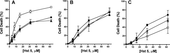
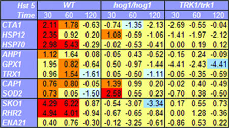
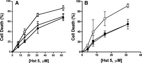
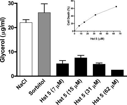
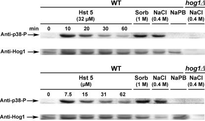
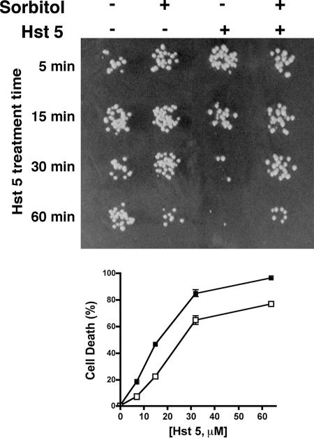
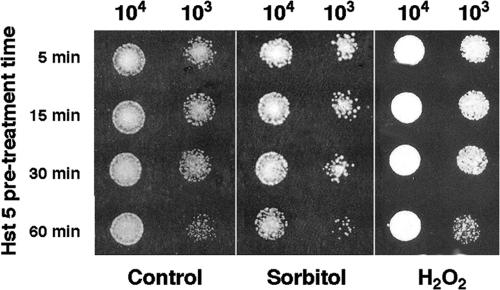
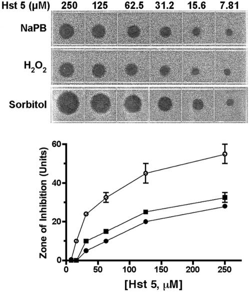
Histatin 5 (Hst 5) is a salivary cationic peptide that has toxicity for Candida albicans by inducing rapid cellular ion imbalance and cell volume loss. Microarray analyses of peptide-treated cells were used to evaluate global gene responses elicited by Hst 5. The major transcriptional response of C. albicans to Hst 5 was expression of genes involved in adaptation to osmotic stress, including production of glycerol (RHR2, SKO1, and PDC11) and the general stress response (CTA1 and HSP70). The oxidative-stress genes AHP1, TRX1, and GPX1 were mildly induced by Hst 5. Cell defense against Hst 5 was dependent on the Hog1 mitogen-activated protein kinase (MAPK) pathway, since C. albicans hog1/hog1 mutants were significantly hypersensitive to Hst 5 but not to Mkc1 MAPK or Cek1 MAPK mutants. Activation of the high-osmolarity glycerol (HOG) pathway was demonstrated by phosphorylation of Hog1 MAPK as well as by glycerol production following Hst 5 treatment in a dose-dependent manner. C. albicans cells prestressed with sorbitol were less sensitive to subsequent Hst 5 treatment; however, cells treated concurrently with osmotic stress and Hst 5 were hypersensitive to Hst 5. In contrast, cells subjected to oxidative stress had no difference in sensitivity to Hst 5. These results suggest a common underlying cellular response to osmotic stress and Hst 5. The HOG stress response pathway likely represents a significant and effective challenge to physiological levels of Hst 5 and other toxic peptides in fungal cells.


Figures

FIG. 1.
FIG. 1.
The HOG MAPK pathway is involved in the protection of C. albicans from Hst 5 toxicity. C. albicans stress response pathways were tested for their abilities to protect cells from Hst 5 killing. (A) The HOG pathway was tested using the wild-type strain RM1000 (filled squares), a hog1/hog1 deletion mutant (open squares), and a HOG1/hog1 restoration strain (filled triangles). (B) The Mkc1 kinase pathway was examined using the wild-type strain CAI-4 (filled squares), an mkc1/mkc1 mutant (open squares), and a single-allele MKC1/mkc1 strain (filled triangles). (C) The Cek1 MAPK pathway was tested using wild type CAI-4 (filled squares), a cek1/cek1 deletion mutant (open squares), and a CEK1/cek1 strain (filled triangles). Cells were treated with 3.8 to 62 μM Hst 5 for 1 h at 30°C, and loss of cell viability was calculated as [1 − (colonies after Hst 5 treatment/colonies after incubation with buffer only)] × 100. Each data point represents the mean ± standard deviation for at least three independent experiments. Only the C. albicans hog1/hog1 strain showed hypersensitivity to Hst 5, showing that Hog1 and/or upstream genes are involved in the reduction of Hst 5 toxicity.
FIG. 2.
FIG. 2.
Hst 5 treatment of C. albicans causes induction of osmotic-stress genes and general stress genes. C. albicans strain CAI-4 (wild type [WT]), strain JC50 (hog1/hog1), and the Hst 5-insensitive strain TK1 (TRK1/trk1) were incubated with 500 μM Hst 5 (30% lethal dose) for 30 min, 60 min, or 120 min, followed by total-RNA isolation and cDNA synthesis (see Materials and Methods). Quantitative RT-PCR analysis was performed for each gene, and the results are shown as n-fold changes in the gene expression levels of Hst 5-treated cells compared to those for control cells treated with buffer only. Values are means from three independent experiments (standard errors are not shown). Genes whose expression was increased more than twofold upon Hst 5 treatment and was significantly different (P < 0.05) from that for untreated cells are shown in red; those whose expression was increased less than twofold but still significantly different (P < 0.05) from that for untreated cells are shown in orange. Genes whose expression was decreased more than twofold upon Hst 5 treatment and was significantly different (P < 0.05) from that for untreated cells are shown in blue; those whose expression was decreased less than twofold but still significantly are shown in light blue. Hst 5 treatment induced significant expression of stress genes (CTA1, HSP70, and HSP12) as well as of genes involved in glycerol accumulation (SKO1 and RHR2) within 30 to 60 min in wild-type C. albicans cells in a Hog1-dependent manner. Oxidative-stress genes (CAP1 and SOD1) were induced by Hst 5 in both wild-type and hog1/hog1 cells.
FIG. 3.
FIG. 3.
C. albicans cells with an impaired oxidative-stress response are more sensitive to high doses of Hst 5. C. albicans oxidative-stress response pathways were tested for their sensitivities to Hst 5 killing. (A) Cells with inactivation of the transcriptional regulatory protein required for oxidative-stress tolerance independent of Hog1 were tested using a wild-type strain (filled squares), a cap1/cap1 deletion mutant (open squares), and a single-allele CAP1/cap1 strain (filled triangles). (B) The Sln1 branch of the HOG pathway was tested by comparing ssk1/ssk1 cells (open squares) with wild-type CAF2-1 (filled squares) and SSK1/ssk1 (filled triangles) cells. Cells were treated with 3.8 to 62 μM Hst 5 for 1 h at 30°C, and loss of cell viability is expressed as [1 − (colonies after Hst 5 treatment/colonies after incubation with buffer only)] × 100. Both C. albicans cap1/cap1 and ssk1/ssk1 strains showed significantly (P < 0.05) more sensitivity to Hst 5 only at higher dosages (31 μM and 62 μM).
FIG. 4.
FIG. 4.
Hst 5 induces dose-dependent intracellular glycerol production in C. albicans. C. albicans CAI-4 (solid bars) cells were incubated with 7.5 to 62 μM Hst 5 for 1 h in diluted dSD. Cells were treated with buffer alone to determine baseline glycerol production as a result of cell manipulation, while cells were treated with 0.5 M NaCl (open bars) or 1 M sorbitol (shaded bars) as positive controls for glycerol production. Glycerol values were normalized to C. albicans cell numbers, and glycerol levels produced in sham-treated cells were used as basal values. Cells were divided for glycerol determination and a conventional candidacidal assay. Hst 5 treatment caused a dose-dependent increase in intracellular glycerol production up to 15 μM; then, at higher Hst 5 doses, glycerol production was reduced in proportion to cell toxicity (inset).
FIG. 5.
FIG. 5.
Hst 5 activates the HOG pathway via Hog1 phosphorylation. C. albicans cells were collected as for glycerol assays in diluted dSD. CAI-4 cell suspensions (2 × 10⁷ cells) were either incubated with Hst 5 (7.5, 15, 31, or 62 μM) for 30 min (lower panel) or with 32 μM Hst 5 for 0, 10, 20, 30, or 60 min (upper panel). Control cells were manipulated in dSD only; cells were treated with 400 mM NaCl or 1 M sorbitol for positive controls. Cells in which Hog1 had been deleted (hog1/hog1) were stimulated with 400 mM NaCl. Following treatment, cells were harvested and directly processed for extraction of total-cell lysates using glass bead disruption. Equal amounts of total-cell lysates (120 μg) were immunoblotted using antibodies to phospho-p38-Hog1p and Hog1p. Hst 5 (32 μM) treatment resulted in rapid phosphorylation of Hog1p after 10 min, which gradually diminished over 60 min (upper panel). Cells treated with buffered medium only (NaPB) showed no Hog1 phosphorylation at any time point. Hog1p phosphorylation was not detected in Hog1 mutants (hog1/hog1). Phosphorylation of Hog1 was strongest when cells were treated with 7.5 or 15 μM Hst 5 (intensity was equivalent in some immunoblots), while cells treated with higher doses of Hst 5 (31 or 62 μM) had reduced levels of Hog1 phosphorylation. WT, wild type.
FIG. 6.
FIG. 6.
Sorbitol-stressed cells are resistant to subsequent treatment with Hst 5. CAF4-2 cells were suspended in diluted dSD (106 cells in 500 μl), exposed to osmotic stress by addition of sorbitol (final concentration, 1 M), and incubated at 30°C for 1 h. Control cells were manipulated in dSD medium alone or with 1 M sorbitol. (Upper panel) To analyze the effects of Hst 5 at various time points, samples were withdrawn after 5, 15, 30, and 60 min of incubation with Hst 5 (7 μM), immediately diluted with buffer, and spotted onto YPD agar plates. Sorbitol protection from Hst 5 cell toxicity was evident following 30 min of incubation with 7 μM peptide (far right), while control cells treated with Hst 5 had typical loss of viability. (Lower panel) To assess concentration effects, cell aliquots were removed, Hst 5 (7 μM to 62 μM) was added, and cells were incubated for 1 h before being plated. Cell death was calculated as described in the legend to Fig. 1. Cells stressed with 1 M sorbitol before Hst 5 addition (open squares) were protected from Hst 5 compared with cells not pretreated with sorbitol (solid squares) at all Hst 5 doses, although the reduction in toxicity was more marked at lower doses.
FIG. 7.
FIG. 7.
Hst 5 pretreatment increases cell sensitivity to subsequent osmotic, but not oxidative, stress. C. albicans (CAF4-2) cells (106/ml) were manipulated as for candidacidal assays. For Hst 5 stress conditioning, Hst 5 (7 μM) was added to cells at 37°C, and samples were withdrawn after 5, 15, 30, or 60 min. Hst 5-treated cells were diluted with NaPB to stop further Hst 5 uptake and then spotted either onto YPD agar plates (control) or onto YPD agar supplemented with 2 mM H2O2 or 1 M sorbitol. The growth of cells under each condition was examined following 24 h of incubation at 37°C. Cells pretreated with Hst 5 and then grown under oxidative-stress conditions (H2O2) (right) had growth profiles identical to those of cells grown in YPD alone (control) (left). Cells pretreated with Hst 5 and then grown under osmotic stress in 1 M sorbitol (center) exhibited increased sensitivity to Hst 5.
FIG. 8.
FIG. 8.
Osmotic stress, but not oxidative stress, increases cell susceptibility to Hst 5. For radial diffusion assays, C. albicans cells (106/ml) were trapped in thin underlay gels, some of which were supplemented with sorbitol (1 M) or H2O2 (1 mM). Twofold dilutions of Hst 5 (7.8 to 250 μM) were loaded into 3-mm-diameter wells that had been punched into underlay gels. After overnight incubation at 37°C, the clear-zone diameters indicating antifungal activity (upper panel) were measured to the nearest 0.1 mm and graphed against peptide concentrations (lower panel). Zone diameters are expressed in units (0.1 mm = 1 U). Cells grown under concurrent osmotic stress (1 M sorbitol) (open circles) were significantly more susceptible to Hst 5 at all concentrations tested than cells grown in medium alone (solid squares), while cells grown under concurrent oxidative stress (1 mM H2O2) (solid circles) showed no difference in sensitivity to Hst 5 (lower panel).

Similar articles

Cited by

References

    1. Alonso-Monge, R., F. Navarro-García, G. Molero, R. Diez-Orejas, J. Pla, M. Sanchez, and C. Nombela. 1999. Role of the mitogen-activated protein kinase Hog1p in morphogenesis and virulence of Candida albicans. J. Bacteriol. 181:3058-3068. - PMC - PubMed
    1. Alonso-Monge, R., F. Navarro-García, E. Román, A. I. Negredo, B. Eisman, C. Nombela, and J. Pla. 2003. The Hog1 mitogen-activated protein kinase is essential in the oxidative stress response and chlamydospore formation in Candida albicans. Eukaryot. Cell 2:351-361. - PMC - PubMed
    1. Arana, D. M., C. Nombela, R. Alonso-Monge, and J. Pla. 2005. The Pbs2 MAP kinase kinase is essential for the oxidative stress response in the fungal pathogen Candida albicans. Microbiology 151:1033-1049. - PubMed
    1. Baev, D., X. S. Li, J. Dong, P. Keng, and M. Edgerton. 2002. Human salivary histatin 5 causes disordered volume regulation and cell cycle arrest in Candida albicans. Infect. Immun. 70:4777-4784. - PMC - PubMed
    1. Baev, D., A. Rivetta, S. Vylkova, J. N. Sun, G. F. Zeng, C. L. Slayman, and M. Edgerton. 2004. The TRK1 potassium transporter is the critical effector for killing of Candida albicans by the cationic protein, histatin 5. J. Biol. Chem. 279:55060-55072. - PubMed

Publication types

MeSH terms