. 2007 Nov 15;110(10):3744-52.
doi: 10.1182/blood-2007-05-093294. Epub 2007 Aug 22.

MLN3897, a novel CCR1 inhibitor, impairs osteoclastogenesis and inhibits the interaction of multiple myeloma cells and osteoclasts

Affiliations

MLN3897, a novel CCR1 inhibitor, impairs osteoclastogenesis and inhibits the interaction of multiple myeloma cells and osteoclasts

Sonia Vallet et al. Blood. .

Abstract

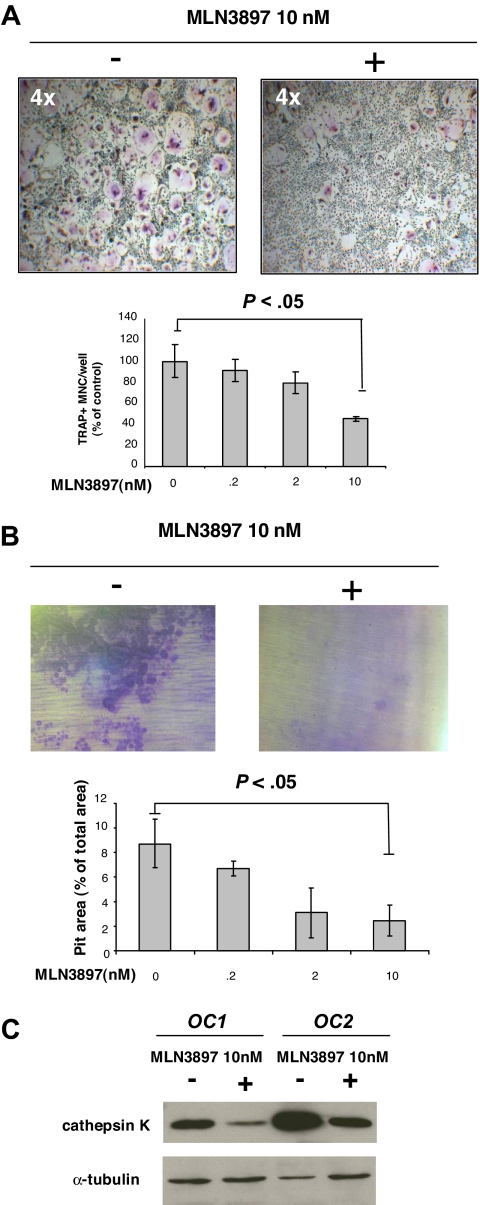
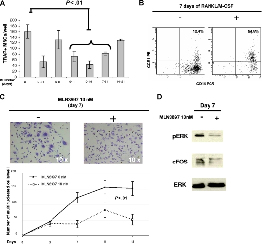
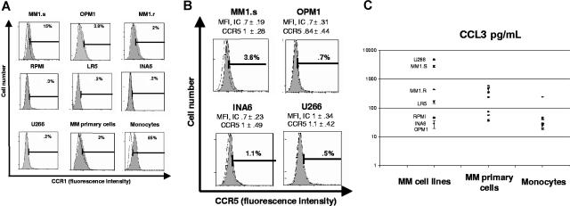
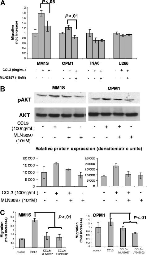
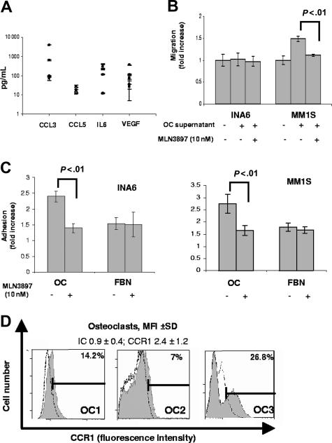
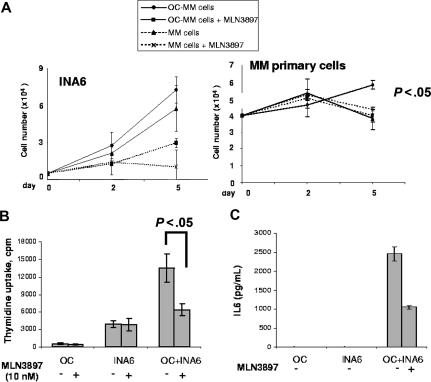
The interaction between osteoclasts (OCs) and multiple myeloma (MM) cells plays a key role in the pathogenesis of MM-related osteolytic bone disease (OBD). MM cells promote OC formation and, in turn, OCs enhance MM cell proliferation. Chemokines are mediators of MM effects on bone and vice versa; in particular, CCL3 enhances OC formation and promotes MM cell migration and survival. Here, we characterize the effects of MLN3897, a novel specific antagonist of the chemokine receptor CCR1, on both OC formation and OC-MM cell interactions. MLN3897 demonstrates significant impairment of OC formation (by 40%) and function (by 70%), associated with decreased precursor cell multinucleation and down-regulation of c-fos signaling. OCs secrete high levels of CCL3, which triggers MM cell migration; conversely, MLN3897 abrogates its effects by inhibiting Akt signaling. Moreover, MM cell-to-OC adhesion was abrogated by MLN3897, thereby inhibiting MM cell survival and proliferation. Our results therefore show novel biologic sequelae of CCL3 and its inhibition in both osteoclastogenesis and MM cell growth, providing the preclinical rationale for clinical trials of MLN3897 to treat OBD in MM.


Figures

Figure 1
Figure 1
MLN3897 inhibits OC formation and function. Adherent PBMCs were cultured for 3 weeks in the presence of RANKL and M-CSF (50 ng/mL) with or without MLN3897 (0.2-10 nM). (A) Cells were stained for TRAP activity. Multinuclear TRAP+ cells are expressed as percentage of control. (B) Adherent PBMCs were cultured on dentine slices. After 3 weeks, resorption areas were stained with toluidine blue, and pit areas were quantified by light microscopy using the public domain NIH Image J program. Each value represents the mean (± SD) of resorptive areas of at least 3 wells, expressed as a percentage of total area. Images were obtained using a Leica DM IL microscope equipped with a 4×, 10×/0.22, and 20×/0.40 numeric aperture objective lens (Leica Microsystems, Wetzlar, Germany) and acquired through IM50 software (Leica Microsystems Imaging Solutions, Cambridge, United Kingdom). All experiments were performed independently at least 3 times. (C) After 3 weeks, we investigated cathepsin K expression on OCs obtained from 2 different donors and treated with MLN3897 (10 nM). Cells were harvested with cell dissociation buffer and lysed; proteins were subjected to immunoblotting with anti–cathepsin K antibody. To ensure equal protein loading, membrane was blotted for tubulin expression.
Figure 2
Figure 2
MLN3897 blocks fusion of OC precursor cells. (A) Adherent PBMCs cultured with RANKL and M-CSF (50 ng/mL) were exposed to MLN3897 (10 nM) for the indicated time points. After 3 weeks, TRAP+ multinucleated cells were stained and counted. Results are expressed as number of TRAP+ multinucleated cells per well: each value represents the mean (± SD) of OCs per well of at least 3 wells. All experiments were performed independently at least 3 times. (B) CCR1 and CD14 coexpression was analyzed by flow cytometry on human PBMCs cultured with or without RANKL and M-CSF (50 ng/mL) for 7 days. (C) PBMCs were cultured in the presence of RANKL and M-CSF with or without MLN3897 (10 nM). At the indicated time points, cells were fixed and stained with hematoxylin/eosin. Cells with more than 3 nuclei were enumerated; images were obtained with a light microscope (Leica Microsystems, Wetzlar, Germany) and acquired through IM50 software (Leica Microsystems Imaging Solutions, Cambridge, United Kingdom). (D) At day 7, PBMCs stimulated with RANKL and M-CSF (50 ng/mL) in the presence or absence of MLN3897 (10 nM) were harvested and lysed; proteins were immunoblotted with anti-pERK, ERK, and c-fos antibodies.
Figure 3
Figure 3
CCR1 expression and CCL3 secretion by MM cell lines and primary MM cells. (A) CCR1 expression on MM cell lines and patient MM cells was analyzed by flow cytometry. A representative flow cytometry shows percentage of positive cells from at least 3 different experiments. CCR1 expression on monocytes is shown as a positive control. (B) CCR5 expression on MM1.S, OPM1, INA6, and U266 was analyzed. Flow cytometry data represent several independent experiments. (C) CCL3 secretion was assessed by ELISA on 48-hour culture supernatants of MM cell lines, MM primary cells, and monocytes plated at a density of 106 cells/mL. Means (± SD) are expressed as pg/mL on a logarithmic scale.
Figure 4
Figure 4
MLN3897 abrogates CCL3-induced migration by inhibiting Akt phosphorylation. Effects of MLN3897 on cell migration in the presence of CCL3 (5 ng/mL) were assessed by Transwell migration assay. MM cells were preincubated with MLN3897 (10 nM for 4 hours) (A) or PI3K inhibitor LY94002 (25 μM for 1 hour) (C), and then seeded on the upper chamber of the Transwell plate. After 4 hours, cells that migrated to the lower chamber were counted, and results were expressed as fold increase over control plus or minus standard deviation. (B) CCL3 (100 ng/mL for 30 minutes) stimulated Akt phosphorylation in serum-starved (overnight in 1% and 30 minutes in 0% RPMI media) MM cells, assessed by Western blot analysis. In selected experiments, cells were preincubated with MLN3897 for 4 hours. The densitometric analysis of the immunoblots displays partial inhibition of Akt phosphorylation by MLN3897 in MM.1S and complete inhibition in OPM1 cells. Results are expressed as mean (± SD).
Figure 5
Figure 5
MLN3897 inhibits MM cell migration and adhesion to OCs. (A) CCL3, CCL5, IL-6, and VEGF secretion was assessed by ELISA in OC supernatants at 48 hours; means (± SD) of 3 independent experiments are expressed as pg/mL on a logarithmic scale. (B) Effects of MLN3897 on cell migration triggered by 48-hour OC supernatants, assessed by Transwell migration assay. INA6 and MM1.S cell lines were preincubated with MLN3897 (10 nM for 4 hours) and seeded in the upper chamber of the Transwell plate. After 4 hours, cells that migrated to the lower chamber were counted. Results are expressed as fold increase over control. (C) Adhesion assay was performed seeding calcein-labeled INA6 and MM1.S cells on OCs (10 000 cells/well) or fibronectin (FBN; 20 μg/mL) in the presence or absence of MLN3897. After 6 hours, nonadherent cells were washed off, and fluorescence intensity of adherent cells were expressed as fold increase over control. (D) Representative CCR1 expression on mature OCs from 3 different donors, analyzed by flow cytometry. Results are expressed as MFI (± SD) of CCR1 and isotype control (IC) from more than 3 independent experiments.
Figure 6
Figure 6
MLN3897 overcomes the survival and proliferation advantage conferred by OCs in coculture. (A) We assessed cell survival of INA6 and MM primary cells alone and in the presence of OCs, with or without MLN3897. At 2 and 5 days, viable cells were counted by trypan blue staining. (B) INA6 cells were cocultured with mature OCs with or without MLN3897 (10 nM) and cell proliferation was assessed by thymidine uptake at 48 hours. Results are expressed as means (± SD) counts per minute (cpm). (C) At the same time point, IL-6 secretion was evaluated by ELISA. Results are expressed as mean (± SD).

References

    1. Body JJ. Effectiveness and cost of bisphosphonate therapy in tumor bone disease. Cancer. 2003;97:859–865. - PubMed
    1. Abe M, Hiura K, Wilde J, et al. Osteoclasts enhance myeloma cell growth and survival via cell-cell contact: a vicious cycle between bone destruction and myeloma expansion. Blood. 2004;104:2484–2491. - PubMed
    1. Charo IF, Ransohoff RM. The many roles of chemokines and chemokine receptors in inflammation. N Engl J Med. 2006;354:610–621. - PubMed
    1. Tian Y, New DC, Yung LY, et al. Differential chemokine activation of CC chemokine receptor 1-regulated pathways: ligand selective activation of Galpha 14-coupled pathways. Eur J Immunol. 2004;34:785–795. - PubMed
    1. Godessart N. Chemokine receptors: attractive targets for drug discovery. Ann N Y Acad Sci. 2005;1051:647–657. - PubMed

Publication types

MeSH terms