

doi: 10.1021/ja074446s.
Epub 2007 Aug 23.
Direct spectroscopic evidence for a high-spin Fe(IV) intermediate in tyrosine hydroxylase
Affiliations
- PMID: 17715926
- PMCID: PMC2860260
- DOI: 10.1021/ja074446s
Item in Clipboard
Direct spectroscopic evidence for a high-spin Fe(IV) intermediate in tyrosine hydroxylase
J Am Chem Soc.
.
No abstract available
Figures

4.2-K Mössbauer spectra of the reaction at 5 °C of the TyrH•Fe(II) •6-MePH4•Tyr complex (2.15 mM TyrH, 1.95 mM Fe(II), 3.7 mM 6-MePH4 and 3.7 mM Tyr in 200 mM Hepes, 10% glycerol, 0.1 M KCl at pH 7.5) with 1.9 mM oxygen-containing buffer in a ratio of 1:2. Reaction times and magnetic field strengths are as indicated. Left panel: Spectra (hashed marks) at various reaction times. The solid lines are quadrupole doublet simulations of the Fe(IV) intermediate (δ = 0.25 mm/s and ΔEQ = 1.27 mm/s). Right panel: Deconvolution of the spectrum of the 20-ms sample in zero-field (top panel) and an 8-T field (bottom panel). The spectrum of the anaerobic control scaled to 60% of the total intensity is shown as a solid line overlaid with the raw data. The resulting difference spectra (hashed marks) can be simulated with spin Hamiltonian simulations of the Fe(IV) intermediate (24% intensity) with the following parameters: S = 2, D = 12.5 cm−1, E/D = 0.05, δ = 0.25 mm/s, ΔEQ = −1.27 mm/s, η = −0.5, A/gNβN = (−18.0, −18.0, −31.0) T.

Comparison of time courses for Fe(IV)O formation and decay (diamonds) and for DOPA formation (circles). DOPA was quantified by rapid-quench of the reaction at 5 °C of the complex of 500 μM TyrH, 480 μM Fe(II), 1 mM Tyr and 2 mM 6-MePH4 with an equal volume of 1.9 mM oxygen-containing buffer. The lines are simulations using the mechanism of Scheme 2 and values of k1 and k2 of 24 and 35 s−1, respectively, assuming that 80% of the enzyme complex is active.

References
-
-
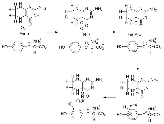
Abbreviations used: TyrH, tyrosine hydroxylase; Tyr, tyrosine; DOPA, dihydroxyphenylalanine; 6-MePH4, 6-methyl tetrahydropterin; 4a-HOPH3, 4a-hydroxypterin.
-
-
- Fitzpatrick PF. Ann Rev Biochem. 1999;68:355–381. - PubMed
-
- Fitzpatrick PF. In: Advances in Enzymology and Related Areas of Molecular Biology. Purich DL, editor. Vol. 74. John Wiley & Sons; New York: 2000. pp. 235–294. - PubMed
-
- Que L., Jr Nat Struct Biol. 2000;7:182–184. - PubMed
-
- Goodwill KE, Sabatier C, Marks C, Raag R, Fitzpatrick PF, Stevens RC. Nat Struct Biol. 1997;4:578–585. - PubMed
Publication types
MeSH terms
Substances
Grants and funding
LinkOut - more resources
Full Text Sources
Other Literature Sources
Medical
