

In vivo micro-CT scanning of a rabbit distal femur: repeatability and reproducibility
- PMID: 17716676
- PMCID: PMC2244798
- DOI: 10.1016/j.jbiomech.2007.06.028
In vivo micro-CT scanning of a rabbit distal femur: repeatability and reproducibility
Abstract
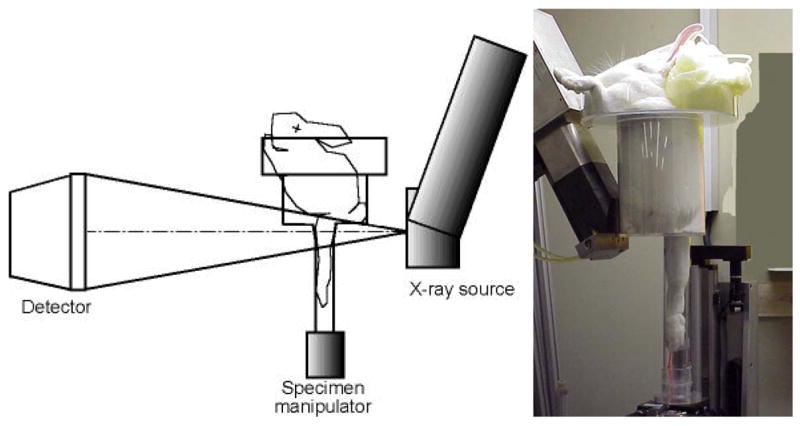
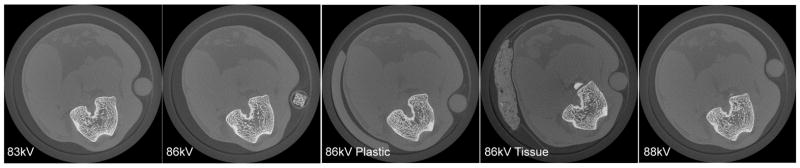
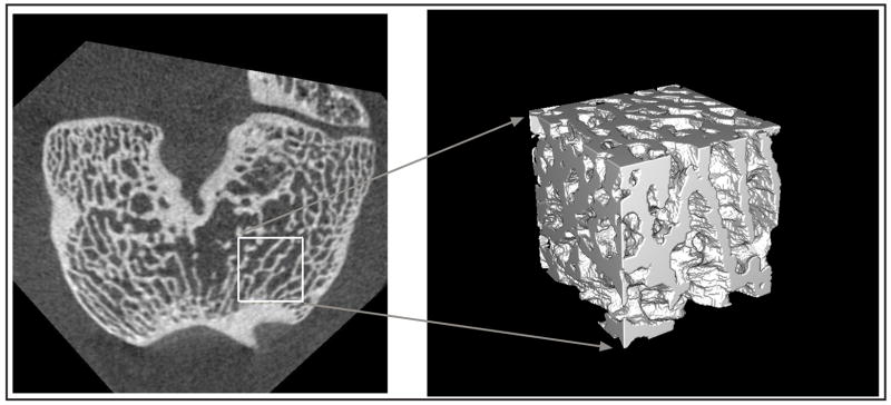
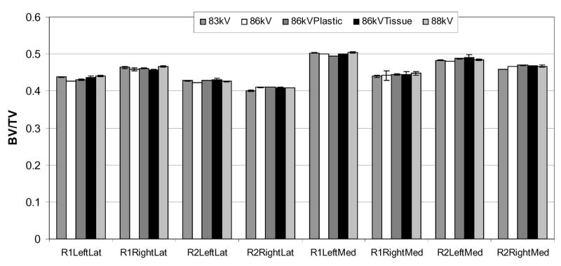
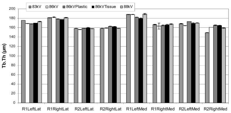
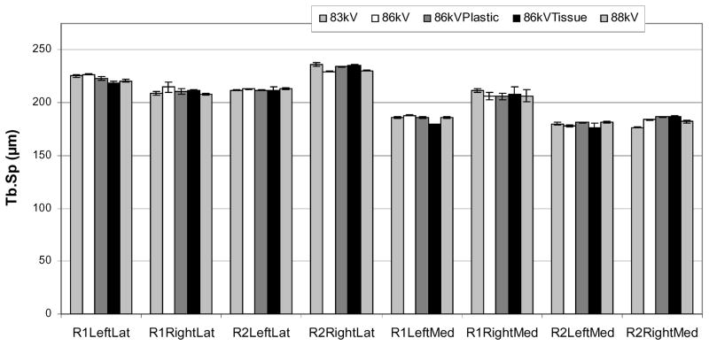
Before in vivo micro-CT scanning can be used to investigate femoral trabecular microarchitecture over time in rabbits, its repeatability and reproducibility must be demonstrated. To accomplish this, both distal femurs of two 6-month-old New Zealand white rabbits were scanned five times each in 1 day under different conditions (repeatability). Scanning was done at 28 microm isotropic voxel size to produce five image stacks of each femur. Three operators then followed a standard image processing protocol (reproducibility) to isolate two separate cubes from each anterior femoral condyle [total n = (8 cube sites)(5 scans)(3 operators) = 120]. Bone volume fraction (BV/TV) of the eight different cube sites (sample) ranged from 0.408 to 0.501 (mean: 0.453); trabecular thickness (Tb.Th) ranged from 158.1 to 185.5 microm (mean: 168.6 microm); and trabecular separation (Tb.Sp) ranged from 179.4 to 233.1 microm (mean: 204.7 microm). Using ANOVA and the variance component method, the total process variation was +/- 14.1% of the mean BV/TV of 0.453. The sample variation was +/- 13.9% (p < 0.001), the repeatability was +/- 2.1% (p < 0.001), and the reproducibility was +/- 0.1% (p > 0.05). Results were similar for Tb.Th and Tb.Sp. Though the contribution due to repeatability was statistically significant for each of the three indices, the natural sample differences were far greater than differences caused by repeated scanning under different conditions or by different operators processing the images. These findings suggest that in vivo micro-CT scanning of rabbit distal femurs was repeatable and reproducible and can be used with confidence to measure differences in trabecular bone microarchitecture at a single location in a longitudinal study design.
Figures
Similar articles
-
Accuracy and precision of segmentation and quantification of wrist bone microarchitecture using photon-counting computed tomography ex vivo.Bone. 2025 May;194:117443. doi: 10.1016/j.bone.2025.117443. Epub 2025 Mar 1. Bone. 2025. PMID: 40032018
-
Validation of calcaneus trabecular microstructure measurements by HR-pQCT.Bone. 2018 Jan;106:69-77. doi: 10.1016/j.bone.2017.09.013. Epub 2017 Oct 3. Bone. 2018. PMID: 28986143
-
Comparison of synchrotron radiation and conventional x-ray microcomputed tomography for assessing trabecular bone microarchitecture of human femoral heads.Med Phys. 2006 Sep;33(9):3568-77. doi: 10.1118/1.2256069. Med Phys. 2006. PMID: 17022253
-
Interindividual and intraspecimen variability of 3-D bone microarchitectural parameters in iliac crest biopsies imaged by conventional micro-computed tomography.J Bone Miner Metab. 2008;26(5):506-13. doi: 10.1007/s00774-008-0856-2. Epub 2008 Aug 30. J Bone Miner Metab. 2008. PMID: 18758910
-
Multiscale and multimodality computed tomography for cortical bone analysis.Phys Med Biol. 2016 Dec 21;61(24):8553-8576. doi: 10.1088/0031-9155/61/24/8553. Epub 2016 Nov 15. Phys Med Biol. 2016. PMID: 27845939
Cited by
-
Cortical Bone Porosity in Rabbit Models of Osteoporosis.J Bone Miner Res. 2020 Nov;35(11):2211-2228. doi: 10.1002/jbmr.4124. Epub 2020 Sep 22. J Bone Miner Res. 2020. PMID: 32614975 Free PMC article.
-
Hindlimb immobilization in a wheelchair alters functional recovery following contusive spinal cord injury in the adult rat.Neurorehabil Neural Repair. 2011 Oct;25(8):729-39. doi: 10.1177/1545968311407519. Epub 2011 Jun 22. Neurorehabil Neural Repair. 2011. PMID: 21697451 Free PMC article.
-
Bone loss following spinal cord injury in a rat model.J Neurotrauma. 2012 May 20;29(8):1676-82. doi: 10.1089/neu.2011.2037. Epub 2012 Feb 22. J Neurotrauma. 2012. PMID: 22181016 Free PMC article.
-
Minimizing Interpolation Bias and Precision Error in In Vivo µCT-Based Measurements of Bone Structure and Dynamics.Ann Biomed Eng. 2016 Aug;44(8):2518-2528. doi: 10.1007/s10439-015-1527-9. Epub 2016 Jan 19. Ann Biomed Eng. 2016. PMID: 26786342 Free PMC article.
-
In vivo monitoring of bone microstructure by propagation-based phase-contrast computed tomography using monochromatic synchrotron light.Lab Invest. 2020 Jan;100(1):72-83. doi: 10.1038/s41374-019-0337-3. Epub 2019 Oct 22. Lab Invest. 2020. PMID: 31641229
References
-
- Bourne BC, Morgan TG, Paschalis EP, Van Der Meullen MC. Cancellous bone anisotropy arises from both architecture and material properties. Proceedings of the 48th Annual Meeting of the Orthopaedic Research Society; Dallas TX. 2002. p. #558.
-
- Bourne BC, van der Meulen MC. Finite element models predict cancellous apparent modulus when tissue modulus is scaled from specimen CT-attenuation. J Biomech. 2004;37(5):613–21. - PubMed
-
- Boyd SK, Davison P, Muller R, Gasser JA. Monitoring individual morphological changes over time in ovariectomized rats by in vivo micro-computed tomography. Bone. 2006a;39(4):854–62. - PubMed
-
- Boyd SK, Moser S, Kuhn M, Klinck RJ, Krauze PL, Muller R, Gasser JA. Evaluation of three-dimensional image registration methodologies for in vivo micro-computed tomography. Ann Biomed Eng. 2006b;34(10):1587–99. - PubMed
-
- Dallari D, Fini M, Stagni C, Torricelli P, Nicoli Aldini N, Giavaresi G, Cenni E, Baldini N, Cenacchi A, Bassi A, Giardino R, Fornasari PM, Giunti A. In vivo study on the healing of bone defects treated with bone marrow stromal cells, platelet-rich plasma, and freeze-dried bone allografts, alone and in combination. J Orthop Res. 2006;24(5):877–88. - PubMed
