

The spinobulbar system in lamprey
- PMID: 17716741
- PMCID: PMC2246055
- DOI: 10.1016/j.brainresrev.2007.07.010
The spinobulbar system in lamprey
Abstract
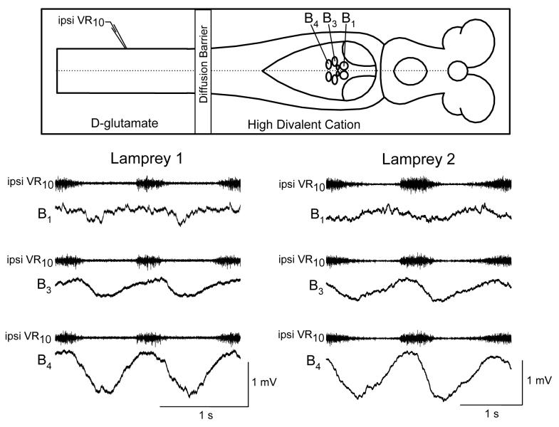
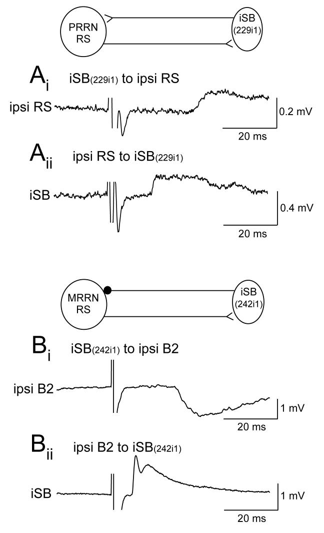
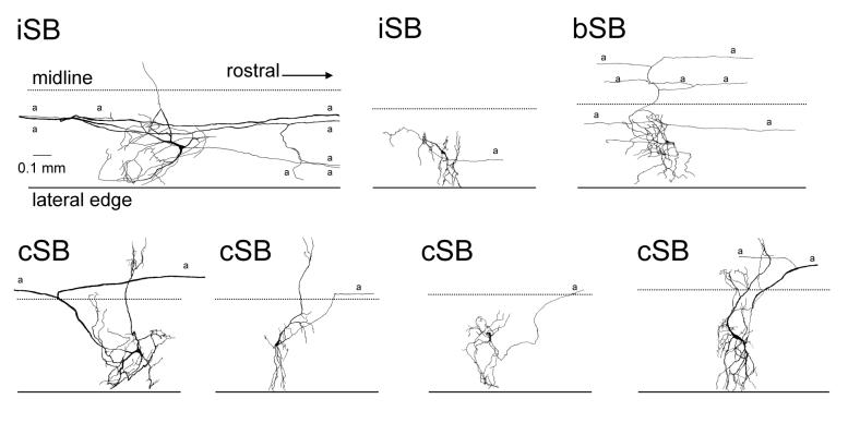
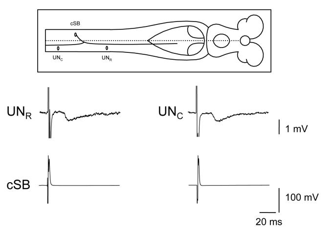
Locomotor networks in the spinal cord are controlled by descending systems which in turn receive feedback signals from ascending systems about the state of the locomotor networks. In lamprey, the ascending system consists of spinobulbar neurons which convey spinal network signals to the two descending systems, the reticulospinal and vestibulospinal neurons. Previous studies showed that spinobulbar neurons consist of both ipsilaterally and contralaterally projecting cells distributed at all rostrocaudal levels of the spinal cord, though most numerous near the obex. The axons of spinobulbar neurons ascend in the ventrolateral spinal cord and brainstem to the caudal mesencephalon and within the dendritic arbors of reticulospinal and vestibulospinal neurons. Compared to mammals, the ascending system in lampreys is more direct, consisting of excitatory and inhibitory monosynaptic inputs from spinobulbar neurons to reticulospinal neurons. The spinobulbar neurons are rhythmically active during fictive locomotion, representing a wide range of timing relationships with nearby ventral root bursts including those in phase, out of phase, and active during burst transitions between opposite ventral roots. The spinobulbar neurons are not simply relay cells because they can have mutual synaptic interactions with their reticulospinal neuron targets and they can have synaptic outputs to other spinal neurons. Spinobulbar neurons not only receive locomotor inputs but also receive direct inputs from primary mechanosensory neurons. Due to the relative simplicity of the lamprey nervous system and motor control system, the spinobulbar neurons and their interactions with reticulospinal neurons may be advantageous for investigating the general organization of ascending systems in the vertebrate.
Figures

Similar articles
-
Spinobulbar neurons in lamprey: cellular properties and synaptic interactions.J Neurophysiol. 2006 Oct;96(4):2042-55. doi: 10.1152/jn.01331.2005. Epub 2006 Jul 12. J Neurophysiol. 2006. PMID: 16837656
-
Spinal locomotor inputs to individually identified reticulospinal neurons in the lamprey.J Neurophysiol. 2011 Nov;106(5):2346-57. doi: 10.1152/jn.01100.2010. Epub 2011 Aug 10. J Neurophysiol. 2011. PMID: 21832033 Free PMC article.
-
Reticulospinal neurons receive direct spinobulbar inputs during locomotor activity in lamprey.J Neurophysiol. 2004 Sep;92(3):1384-90. doi: 10.1152/jn.00625.2003. J Neurophysiol. 2004. PMID: 15331645
-
Initiation of locomotion in lampreys.Brain Res Rev. 2008 Jan;57(1):172-82. doi: 10.1016/j.brainresrev.2007.07.016. Epub 2007 Aug 22. Brain Res Rev. 2008. PMID: 17916380 Review.
-
Flexibility in the patterning and control of axial locomotor networks in lamprey.Integr Comp Biol. 2011 Dec;51(6):869-78. doi: 10.1093/icb/icr077. Epub 2011 Jul 9. Integr Comp Biol. 2011. PMID: 21743089 Free PMC article. Review.
Cited by
-
The contribution of synaptic inputs to sustained depolarizations in reticulospinal neurons.J Neurosci. 2009 Jan 28;29(4):1140-51. doi: 10.1523/JNEUROSCI.3073-08.2009. J Neurosci. 2009. PMID: 19176823 Free PMC article.
-
Organization of an ascending circuit that conveys flight motor state in Drosophila.Curr Biol. 2024 Mar 11;34(5):1059-1075.e5. doi: 10.1016/j.cub.2024.01.071. Epub 2024 Feb 22. Curr Biol. 2024. PMID: 38402616 Free PMC article.
-
Cellular substrates of action selection: a cluster of higher-order descending neurons shapes body posture and locomotion.J Comp Physiol A Neuroethol Sens Neural Behav Physiol. 2008 May;194(5):469-81. doi: 10.1007/s00359-008-0319-1. Epub 2008 Feb 23. J Comp Physiol A Neuroethol Sens Neural Behav Physiol. 2008. PMID: 18297289
-
Remote control of respiratory neural network by spinal locomotor generators.PLoS One. 2014 Feb 20;9(2):e89670. doi: 10.1371/journal.pone.0089670. eCollection 2014. PLoS One. 2014. PMID: 24586951 Free PMC article.
-
Network feedback regulates motor output across a range of modulatory neuron activity.J Neurophysiol. 2016 Jun 1;115(6):3249-63. doi: 10.1152/jn.01112.2015. Epub 2016 Mar 30. J Neurophysiol. 2016. PMID: 27030739 Free PMC article.
References
-
- Arshavsky YI, Berkinblit MB, Fukson OI, Gelfand IM, Orlovsky GN. Recordings of neurones on the dorsal spinocerebellar tract during evoked locomotion. Brain Res. 1972a;43:272–275. - PubMed
-
- Arshavsky YI, Berkinblit MB, Fukson OI, Gelfand IM, Orlovsky GN. Origin of modulation in neurones of the ventral spinocerebellar tract during locomotion. Brain Res. 1972b;43:276–279. - PubMed
-
- Arshavsky YI, Gelfand IM, Orlovsky GN, Pavlova GA. Messages conveyed by spinocerebellar pathways during scratching in the cat. I. Activity of neurons of the lateral reticular nucleus. Brain Res. 1978a;151:479–491. - PubMed
-
- Arshavsky YI, Gelfand IM, Orlovsky GN, Pavlova GA. Messages conveyed by spinocerebellar pathways during scratching in the cat. II. Activity of neurons of the ventral spinocerebellar tract. Brain Res. 1978b;151:493–506. - PubMed
-
- Arshavsky YI, Gelfand IM, Orlovsky GN, Pavlova GA. Messages conveyed by descending tracts during scratching in the cat. I. Activity of vestibulospinal neurons. Brain Res. 1978c;159:99–110. - PubMed
Publication types
MeSH terms
Grants and funding
LinkOut - more resources
Full Text Sources
