. 2007 Sep 4;17(17):1498-503.
doi: 10.1016/j.cub.2007.07.060. Epub 2007 Aug 23.

From stem cell to embryo without centrioles

Affiliations

From stem cell to embryo without centrioles

Naomi R Stevens et al. Curr Biol. .

Abstract

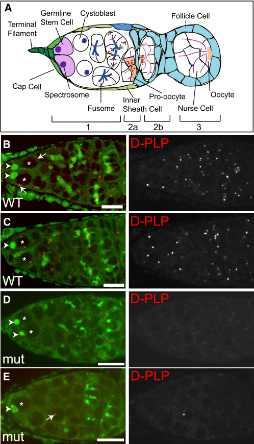
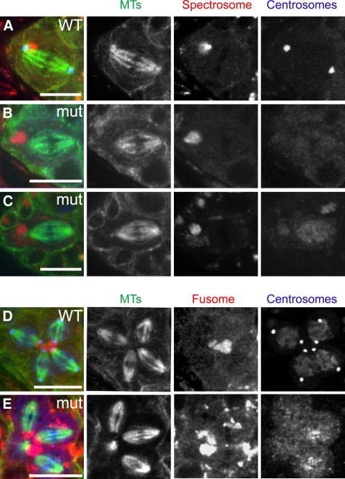
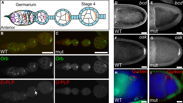
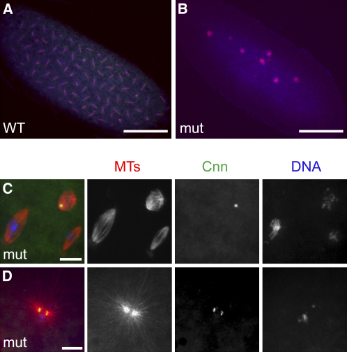
Centrosome asymmetry plays a key role in ensuring the asymmetric division of Drosophila neural stem cells (neuroblasts [NBs]) and male germline stem cells (GSCs) [1-3]. In both cases, one centrosome is anchored close to a specific cortical region during interphase, thus defining the orientation of the spindle during the ensuing mitosis. To test whether asymmetric centrosome behavior is a general feature of stem cells, we have studied female GSCs, which divide asymmetrically, producing another GSC and a cystoblast. The cystoblast then divides and matures into an oocyte, a process in which centrosomes exhibit a series of complex behaviors proposed to play a crucial role in oogenesis [4-6]. We show that the interphase centrosome does not define spindle orientation in female GSCs and that DSas-4 mutant GSCs [7], lacking centrioles and centrosomes, invariably divide asymmetrically to produce cystoblasts that proceed normally through oogenesis-remarkably, oocyte specification, microtubule organization, and mRNA localization are all unperturbed. Mature oocytes can be fertilized, but embryos that cannot support centriole replication arrest very early in development. Thus, centrosomes are dispensable for oogenesis but essential for early embryogenesis. These results reveal that asymmetric centrosome behavior is not an essential feature of stem cell divisions.


Figures

Figure 1
Figure 1
Centrosomes Do Not Segregate Asymmetrically in Female GSC Divisions (A) Schematic diagram of a Drosophila germarium. (B–E) WT (B and C) and DSas-4 mutant (D and E) germaria stained for centrioles (D-PLP [red]) and the spectrosome/fusome (Shot [green]). The nuclei of the GSCs are labeled with an asterisk. In the WT GSCs, neither the single centrosomes (arrows in B) nor the replicated centrosomes (C) adopt a consistent position relative to the spectrosome (arrowheads in B–E) or the stem cell niche. The mutant germarium in (D) has no centrosomes. The germarium in (E) has only a single centrosome (arrow), which is not in a GSC. This demonstrates that the mother centrosome is not selectively retained in female GSCs. Scale bars represent 10 μm.
Figure 2
Figure 2
Centrosomes Are Not Required for Spindle Orientation in Female GSCs or Cysts (A–C) WT (A) and DSas-4 mutant (B and C) female GSCs showing the attachment of one spindle pole to the spectrosome (Shot [red]). Centrosomes, revealed by Cnn (A and B) or Polo (C) staining (blue), are absent in the mutant GSCs. MTs are shown in green. Anterior is to the left. (D–E) WT (D) and DSas-4 mutant (E) four-cell cysts. Despite the absence of centrosomes (revealed by Polo staining in blue) in the mutant cyst, the four spindles (one of which is only partially visible in this Z section) are clearly attached to the fusome. Scale bars represent 10 μm.
Figure 3
Figure 3
The Oocyte Is Correctly Specified and mRNAs Are Properly Localized in the Absence of Centrioles (A) Schematic diagram of Orb localization (light orange) in a Drosophila ovariole. (B and C) WT (B) and DSas-4 mutant (C) ovarioles showing the accumulation of Orb protein (green) in the oocyte of each cyst. D-PLP staining (red) shows the presence of many centrioles in the WT ovariole, and these can be seen to accumulate in the oocyte (arrow). No centrioles are detectable in the mutant ovariole. Scale bars represent 50 μm. (D–G) Stage 10A oocytes showing the localization of bicoid (bcd) mRNA to the anterior margin (D and E) and oskar (osk) mRNA to the posterior pole (F and G). The localization of both mRNAs is indistinguishable in WT oocytes (D and F) and DSas-4 mutant germline clones (E and G). Scale bars represent 25 μm. (H and I) Stage 8 egg chambers showing the localization of Gurken protein (red) between the nucleus and the anterior-dorsal corner of the oocyte. Gurken localizes identically in WT (H) and in a DSas-4 mutant germline clone (I) (marked by the absence of nuclear GFP in green). DNA is in blue. Scale bars represent 20 μm.
Figure 4
Figure 4
Centrioles Are Essential for Early Embryonic Development (A and B) WT (A) and DSas-4 mutant (B) embryos from a 1–4 hr collection stained for tubulin (red), centrosomes (Cnn [green]) and DNA (blue). The WT embryo is ∼1 hr old and contains many well-organized spindles. The mutant embryo, although at least 1 hr old, is arrested early in development with only 6–7 disorganized anastral spindles. Scale bars represent 100 μm. (C) High magnification view of spindles in a DSas-4 mutant embryo. All the spindles appear to be anastral, although one possesses a Cnn dot at one pole. This embryo contained a second Cnn dot not shown here. Scale bars represent 10 μm. (D) A DSas-4 mutant embryo containing two closely associated Cnn dots that each nucleate a large MT aster. Scale bars represent 10 μm.

References

    1. Rebollo E., Sampaio P., Januschke J., Llamazares S., Varmark H., Gonzalez C. Functionally unequal centrosomes drive spindle orientation in asymmetrically dividing Drosophila neural stem cells. Dev. Cell. 2007;12:467–474. - PubMed
    1. Yamashita Y.M., Mahowald A.P., Perlin J.R., Fuller M.T. Asymmetric inheritance of mother versus daughter centrosome in stem cell division. Science. 2007;315:518–521. - PMC - PubMed
    1. Rusan N.M., Peifer M. A role for a novel centrosome cycle in asymmetric cell division. J. Cell Biol. 2007;177:13–20. - PMC - PubMed
    1. McGrail M., Hays T.S. The microtubule motor cytoplasmic dynein is required for spindle orientation during germline cell divisions and oocyte differentiation in Drosophila. Development. 1997;124:2409–2419. - PubMed
    1. Theurkauf W.E. Microtubules and cytoplasm organization during Drosophila oogenesis. Dev. Biol. 1994;165:352–360. - PubMed

Publication types