. 2007 Oct;171(4):1113-26.
doi: 10.2353/ajpath.2007.070226. Epub 2007 Aug 23.

Inhibitor of differentiation 1 promotes endothelial survival in a bleomycin model of lung injury in mice

Affiliations

Inhibitor of differentiation 1 promotes endothelial survival in a bleomycin model of lung injury in mice

Huimin Zhang et al. Am J Pathol. 2007 Oct.

Abstract

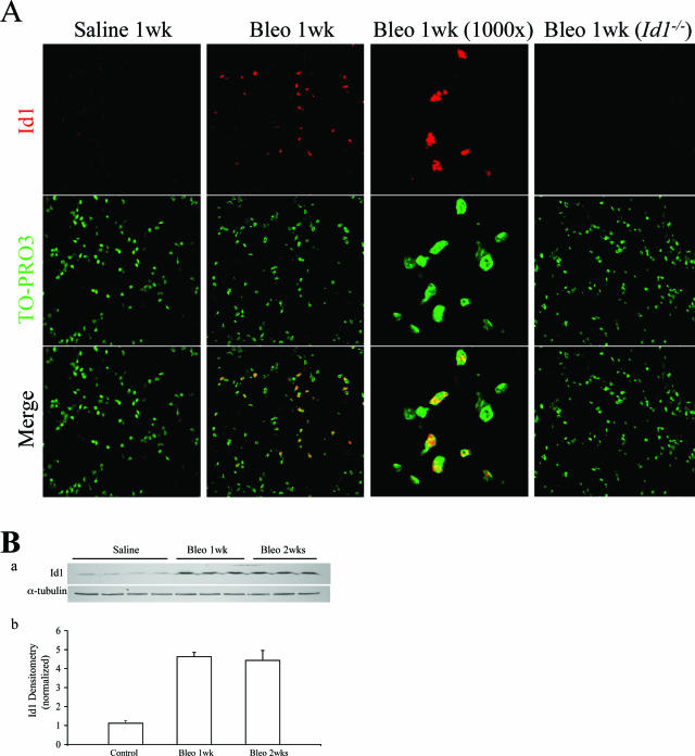
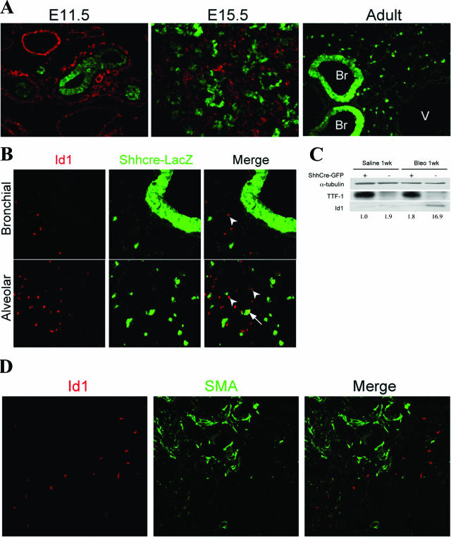
The Id family of genes encodes negative regulators of basic helix-loop-helix transcription factors and has been implicated in diverse cellular processes such as proliferation, apoptosis, differentiation, and migration. However, the specific role of Id1 in lung injury has not been investigated. Bleomycin has been widely used to generate animal models of acute lung injury and fibrogenesis. In this study we found that, on bleomycin challenge, Id1 expression was significantly up-regulated in the lungs, predominantly in endothelial cells, as revealed by double immunolabeling and quantitative flow cytometric analysis. Mice with Id1 loss-of-function (Id1(-/-)) displayed increased vascular permeability and endothelial apoptosis in the lungs after bleomycin-induced injury. Cultured Id1(-/-) lung microvascular endothelial cells also showed decreased survival when exposed to bleomycin. We detected a decrease in the level of Bcl-2, a primary anti-apoptotic protein, in Id1(-/-) endothelial cells, suggesting that down-regulated Bcl-2 may promote endothelial apoptosis in the lung. Therefore, we propose that Id1 plays a crucial role in promoting endothelial survival in the adult lung on injury. In addition, bleomycin-exposed Id1(-/-) mice showed increased lung collagen accumulation and fibrogenesis, suggesting that Id1 up-regulation in the lung may play a critical role in lung homeostasis.


Figures

Figure 1
Figure 1
Id1 expression is significantly up-regulated in the adult lung on bleomycin injury. A: Representative photographs showing sections from 8-week-old wild-type and Id1−/− lungs treated with saline or 0.08 U of bleomycin and collected after 1 week. Sections were immunostained with Id1 (red), and nuclei were counterstained with TO-PRO3 (green). Note the nuclear Id1 expression in wild-type lungs treated with bleomycin but not in saline controls. The specificity of Id1 staining is confirmed by using bleomycin-treated Id1−/− lungs as negative control. B: a: Protein extracts from 8-week-old C57BL/6 wild-type lungs or Id1−/− lungs treated with saline or 0.08 U bleomycin and collected after 1 and 2 weeks were immunoblotted with Id1 antibody. α-Tubulin was used as a loading control. b: Densitometric measurements of Id1 bands were performed and normalized to the density of α-tubulin bands. Original magnifications, ×400.
Figure 2
Figure 2
Id1 expression is up-regulated prominently in the injured lung endothelium. A: Representative photographs showing sections from 8-week-old wild-type lungs at 1 and 2 weeks after bleomycin. Sections were double-stained with Id1 (red) and endothelial marker CD34 (green) antibodies. Arrows in the figures point to representative Id1-expressing endothelial cells; note the membrane and cytoplasmic CD34 staining that wraps around the nuclear Id1 staining. Arrowheads point to Id1-expressing nonendothelial cells. B: Eight-week-old wild-type Tie1Cre-ZEG lungs were treated with saline or 0.08 U of bleomycin and collected after 1 week; lung cells were sorted into endothelial (GFP+) and nonendothelial (GFP) group by FACS. Protein extracts were immunoblotted with Id1 and α-tubulin as the loading control. The endothelial fraction was confirmed by immunoblotting with endothelial marker, PECAM-1. Densitometric measurements of Id1 bands were performed and normalized to the density of α-tubulin bands and numeric values indicating differential levels of expression are shown at the bottom. Note the dramatic up-regulation of Id1 protein level in endothelial cells from bleomycin-treated lungs compared with the nonendothelial fraction and saline controls. C: Eight-week-old wild-type Tie1Cre-ZEG lungs were collected at 2 weeks after bleomycin or saline and then subjected to FACS sorting and Western blotting. Protein extracts were immunoblotted with Id1 and α-tubulin. Densitometric measurements of Id1 bands were performed and normalized to the density of α-tubulin bands. Original magnifications: ×400 (A, low magnification); ×1000 (A, high magnification).
Figure 3
Figure 3
Id1 expression is not significantly up-regulated in bleomycin-exposed lung fibroblasts and ShhCre-GFP-marked epithelium. A: Distribution of GFP-labeled epithelial cells (green) in embryonic lungs at E11.5 and E15.5 and in the adult lung at 6 weeks. Embryonic lung sections were double-stained with endothelial marker PECAM-1 (red). Note that GFP expression is strictly confined in epithelial cells throughout development and is excluded from the PECAM-1-positive endothelial domain. GFP expression was detected in the adult bronchial epithelium (Br) and alveolar epithelial cells. Vessel (V) was negative. B: Representative photographs showing sections from 8-week-old wild-type ShhCre-R26R lungs at 1 week after bleomycin. Sections were double-stained with Id1 (red) and epithelial marker Shhcre-LacZ (green). Arrows in the figures point to representative Id1-expressing epithelial cells. Arrowheads point to Id1-expressing nonepithelial cells. C: Eight-week-old wild-type ShhCre-ZEG lungs collected at 1 week after bleomycin or saline were subjected to FACS sorting and Western blotting. Protein extracts were immunoblotted with Id1 and α-tubulin. Densitometric measurements of Id1 bands were performed and normalized to the density of α-tubulin bands. The epithelial fraction was confirmed by immunoblotting with epithelial marker, TTF-1. D: Representative photographs showing sections from 8-week-old wild-type lungs at 2 weeks after bleomycin. Sections were double-stained with Id1 (red) and myofibroblast marker, smooth muscle α-actin (SMA) (green). Original magnifications: ×200 (A); ×600 (B); ×400 (D).
Figure 4
Figure 4
Bleomycin-injured Id1−/− lungs display increased endothelial barrier dysfunction and elevated endothelial apoptosis. A: Increased vascular permeability in bleomycin-challenged Id1−/− lungs. Pulmonary vascular permeability of saline or bleomycin-treated 8-week-old wild-type or Id1−/− lungs was determined by Evans blue extravasation assay (n = 6 for each group). Note 58% increase in Evans Blue dye leakage in injured Id1−/− lungs compared with wild-type lungs. Asterisks denote a significant difference (P < 0.001) between wild-type and Id1−/− lungs at 1 week after bleomycin. B: Endothelial cell death detection in wild-type and Id1−/− lungs by TUNEL (red) and CD34 (green) double-labeling. Representative sections are shown for lungs at 1 week after bleomycin or saline. C: Flow cytometric counting of apoptotic endothelial cells in wild-type or Id1−/− Tie1Cre;ZEG lungs at 1 week after bleomycin or saline. a: Representative dotspot graphs showing cell sorting results of 10,000 lung cells, using annexin-5 as the cell death marker and GFP as the endothelial marker. The top right quadrant represents apoptotic endothelial cells. b: Statistical representation of the sorting results from six independent experiments (n = 6). Note there is an average 37.6% increase in endothelial cell death in injured Id1−/− lungs compared with wild-type lungs. Asterisks denote a significant difference (P < 0.05) between wild-type and Id1−/− lungs. Original magnifications, ×400.
Figure 5
Figure 5
Id1−/− lung microvascular endothelial cells display reduced survival in culture. A: Id1 is up-regulated in endothelial cells by bleomycin in vitro. Western blotting showing Id1 level from wild-type primary LMVECs treated with saline or 250 ng/ml bleomycin for the indicated time points. Values represent relative fold change of Id1 protein level normalized to the density of α-tubulin bands. B: Cell death detection of wild-type and Id1−/− primary LMVECs by TUNEL (red) and Tie1Cre-GFP (green) double labeling. Representative photographs are shown for cell cultures treated with 250 ng/ml bleomycin for 6 hours followed by no bleomycin for 3 hours. C: Flow cytometric counting of apoptotic endothelial cells in wild-type or Id1−/− Tie1Cre-ZEG LMVECs treated with saline or 250 ng/ml bleomycin for 6 hours. a: Representative dotspot graphs showing the results of cell sorting, at 10,000 cells, each using annexin-5 as the cell death marker and GFP as the endothelial marker. The top right quadrant represents apoptotic endothelial cells. b: Statistical representation of the sorting results from six independent experiments (n = 6). Note that there is nearly twofold increase in endothelial cell death in Id1−/− LMVECs compared with wild type. Asterisks denote a significant difference (P < 0.05) between wild-type and Id1−/− cells. Original magnifications, ×100.
Figure 6
Figure 6
Id1−/− lung microvascular endothelial cells show reduced Bcl-2 level and MEK/ERK activity. Western blotting of cell lysates collected from wild-type or Id1−/− primary LMVECs treated with 250 ng/ml bleomycin for or 12 hours. Note that Bcl-2 level is significantly decreased in LMVECs in the absence of Id1 function. The MEK/ERK signaling pathway is also affected in Id1−/− endothelial cells, based on lower levels of phosphorylated MEK/ERK.
Figure 7
Figure 7
Id1−/− lungs are more susceptible to bleomycin-induced fibrogenesis. A: H&E staining of lung sections to show architectural changes. Representative photographs showing foci formation in wild-type and Id1−/− lungs at 2 weeks after bleomycin. B: Increase in the index of architectural distortion in Id1−/− lungs compared with wild-type lungs after bleomycin challenge. Double asterisks denote a significant difference (P < 0.001) between wild-type and Id1−/− lungs at 2 weeks after bleomycin (n = 9 for each group). C: Trichrome staining showing increased collagen deposition in lungs of Id1−/− mice after bleomycin challenge. Representative photographs reveal increased collagen accumulation (blue) in Id1−/− lungs compared with wild type at 2 weeks after bleomycin. D: Quantification of collagen deposition in lungs of wild-type and Id1−/− mice after bleomycin challenge by hydroxyproline assay. Asterisks denote a significant difference (P < 0.01) between wild-type and Id1−/− lungs at 2 weeks after bleomycin (n = 8 for each group). E: Immunostaining of wild-type and Id1−/− lungs with antibodies specific for α-SMA. Representative sections are shown for lungs at 2 weeks after bleomycin or saline. Sections were counterstained with hematoxylin to highlight all nuclei. Note elevated expression of α-SMA in fibrotic foci of Id1−/− lungs compared with wild type. F: Western blotting showing expression level of α-SMA in wild-type and Id1−/− lungs at 2 weeks after bleomycin or saline. Values represent the relative fold change of α-SMA protein level normalized to the density of α-tubulin bands. Original magnifications, ×400.
Figure 8
Figure 8
Schematic representation of Id1 function in the lung endothelium in acute lung injury. Injury caused by bleomycin results in extensive endothelial apoptosis. The lung responds to tissue damage by up-regulating Id1 expression in endothelial cells (and other cell types). Up-regulation of Id1 in endothelial cells activates the MEK/ERK pathway and elevates expression of anti-apoptotic protein Bcl-2. Increased Bcl-2 protein level reduces the extent of endothelial cell apoptosis, thus alleviating endothelial damage. Acute lung injury often results in pulmonary fibrosis in the chronic phase, which may be attenuated by Id1 function.

Similar articles

Cited by

References

    1. Ruzinova MB, Benezra R. Id proteins in development, cell cycle and cancer. Trends Cell Biol. 2003;13:410–418. - PubMed
    1. Sun XH, Copeland NG, Jenkins NA, Baltimore D. Id proteins Id1 and Id2 selectively inhibit DNA binding by one class of helix-loop-helix proteins. Mol Cell Biol. 1991;11:5603–5611. - PMC - PubMed
    1. Zebedee Z, Hara E. Id proteins in cell cycle control and cellular senescence. Oncogene. 2001;20:8317–8325. - PubMed
    1. Benezra R, Rafii S, Lyden D. The Id proteins and angiogenesis. Oncogene. 2001;20:8334–8341. - PubMed
    1. Wong YC, Wang X, Ling MT. Id-1 expression and cell survival. Apoptosis. 2004;9:279–289. - PubMed

Publication types

MeSH terms