. 2007 Sep 5;26(17):3981-92.
doi: 10.1038/sj.emboj.7601820. Epub 2007 Aug 23.

The specificity of SNARE pairing in biological membranes is mediated by both proof-reading and spatial segregation

Affiliations

The specificity of SNARE pairing in biological membranes is mediated by both proof-reading and spatial segregation

Ioanna Bethani et al. EMBO J. .

Abstract

Soluble N-ethylmaleimide-sensitive factor attachment receptor (SNARE) proteins mediate organelle fusion in the secretory pathway. Different fusion steps are catalyzed by specific sets of SNARE proteins. Here we have used the SNAREs mediating the fusion of early endosomes and exocytosis, respectively, to investigate how pairing specificity is achieved. Although both sets of SNAREs promiscuously assemble in vitro, there is no functional crosstalk. We now show that they not only colocalize to overlapping microdomains in the membrane of early endosomes of neuroendocrine cells, but also form cis-complexes promiscuously, with the proportion of the different complexes being primarily dependent on mass action. Addition of soluble SNARE molecules onto native membranes revealed preference for cognate SNAREs. Furthermore, we found that SNAREs are laterally segregated at endosome contact sites, with the exocytotic synaptobrevin being depleted. We conclude that specificity in endosome fusion is mediated by the following two synergistically operating mechanisms: (i) preference for the cognate SNARE in 'trans' interactions and (ii) lateral segregation of SNAREs, leading to relative enrichment of the cognate ones at the prospective fusion sites.


Figures
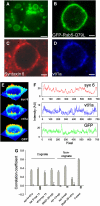
Figure 1
Figure 1
The cognate and non-cognate SNAREs largely colocalize in microdomains on the endosomal membrane. (A) PC12 cells were transfected with a plasmid expressing GFP-Rab5-Q79L. Forty-eight hours post-transfection, cells were fixed and imaged by use of a Zeiss Axiovert 200M fluorescence microscope. The GFP-bound Rab5 variant was observed on vesicular structures of 1–5 μm, which correspond to enlarged early endosomes. Scale bar, 5 μm. (B–D) PC12 cells expressing the GFP-Rab5-Q79L (green) were stained for syntaxin 6 (red channel) and vti1a (blue channel), and imaged by confocal fluorescence microscopy. Images show a typical endosome. Note the SNARE domains on the endosomal membrane. Scale bar, 1 μm. (E) The intensity images in panels B–D are plotted as surfaces, in pseudocolor. Note that the SNAREs are found in domains, which largely correlate. (F) Intensity profiles of syntaxin 6 (red), vti1a (blue) and GFP-Rab5-Q79L (green) signals along the endosomal membrane of the endosome shown in panels B–E. (G) Correlation between the staining of different pairs of SNAREs on the endosomal membrane. Control, costaining of single SNAREs with Cy3- and Cy5-conjugated secondary antibodies. Values are means±s.e. of three independent experiments with 10–15 analyzed endosomes. The striped bars correspond to the negative control (see Materials and methods).
Figure 2
Figure 2
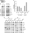
Interactions between non-cognate SNAREs in early endosomes. (A) Enrichment of an early endosomal-specific marker (Rab5) in the fraction isolated from the gradient. Equal amounts of the endosomal fraction (lane1) and post nuclear supernatant (PNS) (lane 2) were analyzed by SDS–PAGE and blotted with anti-Rab5 antibody. For comparison, actin and the NMDA receptor (as a cellular membrane marker) are also shown; the enrichment of endosomes (Rab5 enrichment) was approximately 9.6-fold, on average (±3.9, four experiments). (B) Synaptobrevin interacts with the early endosomal SNAREs. PNS from PC12 cells was centrifuged in a sucrose density gradient, a band highly enriched in early endosomes was isolated, and after solubilization with Triton X-100 immunoprecipitation was performed with a monoclonal antibody against synaptobrevin (left lane); controls are shown in the second lane (no antibody added). The corresponding supernatants (Sup) are shown in the respective position (lanes 3–4). The interacting SNAREs were identified by immunoblot analysis. A total of 10% of the total sample is loaded in every lane. Typical blots of three independent experiments are shown. (C) Quantification by densitometry of coprecipitation, from the blots presented in panel B; the band intensities were normalized to starting material (see Materials and methods). The ‘no antibody' value corresponds to the mean value of all negative controls presented. (D) Other SNARE interactions in the early endosomal fraction. Immunoprecipitations were performed with antibodies against the SNAREs indicated at the top of the figure. All immunoprecipitates were analyzed by immunoblotting for the SNAREs indicated. The ‘no ab' value corresponds to the negative control, in which no antibody was added for immunoprecipitation. A total of 10% of the total sample is loaded in every lane. Boxed bands indicate immunoprecipitation of the blotted protein with its own antibody. Empty arrowheads indicate cognate interactions; full arrowheads indicate non-cognate interactions. Note that SNAP-25 was omitted from the analysis because none of the available antibodies detects SNAP-25 in assembled SNARE complexes. Immunoprecipitation of syntaxin 13 by vti1a and syntaxin 1 is presented from PNS material; identical results were obtained with the endosomal fraction (data not shown). See Supplementary Table 1 for the immunoprecipitation efficiencies of all antibodies.
Figure 3
Figure 3
The non-cognate SNARE interactions represent genuine SNARE complexes in endosomal membranes. (A) PNSs were incubated with rat brain cytosol in presence or absence of ATP, and immunoprecipitations were performed after detergent extraction. Typical immunoblots for syntaxin 13 are shown. Note the presence of bands in the synaptobrevin precipitants in absence, but not in presence of ATP (arrowheads). (B) Quantification of syntaxin 13 co-immunoprecipitation. Bands were quantified by densitometry (see Materials and methods); averages±s.e.m. from three independent experiments are shown. (C) The interaction between synaptobrevin and syntaxin 13 is detectable in absence of NSF activity (arrowhead). PNS fractions were incubated in presence of ATP, cytosol and NEM, to block NSF activity.
Figure 4
Figure 4
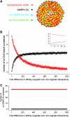
Mass action determines cis-complex formation. We simulated the behavior of SNAREs in a model endosome, containing synaptobrevin, VAMP4, the early endosomal Q-SNARE acceptor complex (syntaxin 13/vti1a/syntaxin 6) and the exocytotic Q-SNARE acceptor complex (syntaxin 1/SNAP-25). We allowed the SNAREs to mix and interact for 1000 iterations, and then counted the number of both cognate and non-cognate complexes. We varied the affinity of R-SNAREs for their non-cognate complex acceptors between 1 and 1/1000. The affinity for the cognate complex was always 1. The fold difference in affinity is indicated on the x-axis. (A) The levels of the different elements are shown in brackets; the graphic description of the model is provided to give an impression of the different SNARE densities. (B) The number of complexes containing the early endosomal Q-SNARE acceptor (synaptobrevin-containing complexes, red; VAMP4-containing complexes, black). As synaptobrevin is the non-cognate partner here, its affinity decreases from left to right. The inset zooms on the first 20 points of the curves. Note that the number of synaptobrevin-containing complexes largely outnumbers that of VAMP4-containing complexes when the affinity difference is of 1–50 fold. Some non-cognate complexes form even at 1000-fold differences in affinity, when VAMP4 is still limited to only 4 of the 5 possible complexes (see also Supplementary Figure 4). (C) The number of complexes containing the exocytotic Q-SNARE acceptor. As VAMP4 is the non-cognate partner here, its affinity decreases from left to right. Note that, due to the differences in concentration, essentially no VAMP4-containing (non-cognate) complexes form, even when the affinity is 1 for both the R-SNAREs (the left-most point).
Figure 5
Figure 5
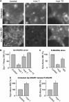
Exocytotic SNAREs bind relatively specifically to plasma membrane sheets. (A) Membrane sheets were treated with 1 μM syntaxin 1A-Alexa 594 alone (left), or in the presence of excess (10 μM) syntaxin 1 (middle) or syntaxin 13 (right). The top three panels show the plasma membranes (by staining with the lipid tracer TMA-DPH). Alexa 594 incorporation in the membrane is shown in the bottom panels. (B) Quantification of syntaxin 1A-Alexa 594 binding; 10-fold excess unlabeled competing SNAREs were added as indicated. (C) Similar experiments were performed to investigate synaptobrevin binding to membrane sheets. Unlabeled synaptobrevin or endobrevin were added as 10-fold excess, as indicated. (D) Membrane sheets were reacted with Alexa 488-labeled R-SNAREs, either alone or in the presence of equimolar amounts (4 μM) of syntaxin 1. The graph on the left shows the results for synaptobrevin; the graph on the right indicates the results for endobrevin.
Figure 6
Figure 6
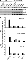
Promiscuous SNARE complexes form between membrane-resident SNAREs and SNAREs added externally at high concentrations. PNS of PC12 cells was incubated with 25 μM of myc-tagged proteins synaptobrevin, VAMP4 and endobrevin, respectively, in presence of ATP at 37°C. After 45 min of incubation, 1 mM NEM was added to block disassembly and incubation continued for 30 min. Reactions were centrifuged for 25 min at 300 000 g, to eliminate the soluble SNAREs molecules not bound to the membranes. Pellets were resuspended in extraction buffer and immunoprecipitated with anti-myc antibodies. Each immunoprecipitation was accompanied by a negative control in which no antibody was used. The precipitants were analyzed by SDS–PAGE Western blotting. We present the results for four SNAREs for which co-immunoprecipitation bands were observed: SNAP-25, syntaxin 1, syntaxin 6 and syntaxin 13. (A) Typical blots are presented. (B–D) The amounts of protein coprecipitating with myc-synaptobrevin (B), myc-VAMP4 (C) and myc-endobrevin (D) were quantified; averages±s.e.m. from four independent experiments are shown (black bars). Gray bars indicate the amount of SNARE complexes that formed after solubilization (see Materials and methods for details). Three independent experiments were performed in conditions identical to those of the experiments in panels A–D; bars show average.
Figure 7
Figure 7
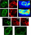
Synaptobrevin appears to avoid the interfaces between endosomes. PC12 cells were transfected with a plasmid expressing GFP-Rab5-Q79L and immunostained for synaptobrevin as in Figure 1. (A) Two endosome pairs showing clear interfaces; green, Rab5-GFP; red, synaptobrevin. Scale bar, 1 μm. (B) Three-dimensional view of the endosomes in the top panels of A. Top, Rab5-GFP. Bottom, synaptobrevin. Arrows point to the interface. (C) Endosome pair similarly stained for synaptobrevin (middle) and syntaxin 6 (right). (D) The fate of the endosomes pressing against each other is difficult to predict; they most likely fuse, as we see relatively abundant constricted (i.e. post-fusion) structures.
Figure 8
Figure 8
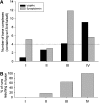
Fusion specificity is achieved by synergistically operating mechanisms. We simulated the behavior of two interacting endosomes of ∼233-nm diameter, each containing 25 VAMP4 molecules, 40 early endosomal Q-SNARE acceptor complexes (syntaxin 13/vti1a/syntaxin 6), 2195 synaptobrevin molecules and 170 exocytotic Q-SNARE acceptor complexes (syntaxin 1/SNAP-25). We allowed the SNAREs from the two endosomes to interact in the interface between them for 1000 iterations. All trans-complexes that formed remained stable until the end of simulation; cis-complexes formed as well, but were always disengaged before the start of the next iteration. (A) We monitored the number of trans-complexes containing the early endosomal Q-SNAREs (with either VAMP4, shown in black, or synaptobrevin, shown in gray). A 10-fold preference for cognate complex formation was modeled (see Supplementary Figure 6 for other preference values). Four models are presented as follows: I, random distribution of both exocytotic and endosomal SNAREs; II, 10-fold de-enrichment of exocytotic SNAREs from the interface; III, three-fold enrichment of endosomal SNAREs in the interface and IV, both a de-enrichment of exocytotic SNAREs and an enrichment of the endosomal ones. (B) Formation of at least four VAMP4-containing complexes was required to consider the two endosomes as ‘fused'. The average percentage of fused pairs for 30–45 simulations is shown.

References

    1. Aikawa Y, Lynch KL, Boswell KL, Martin TF (2006) A second SNARE role for exocytic SNAP25 in endosome fusion. Mol Biol Cell 17: 2113–2124 - PMC - PubMed
    1. Antonin W, Riedel D, Fischer von Mollard G (2000) The SNARE Vti1a-beta is localized to small synaptic vesicles and participates in a novel SNARE complex. J Neurosci 20: 5724–5732 - PMC - PubMed
    1. Brandhorst D, Zwilling D, Rizzoli SO, Lippert U, Lang T, Jahn R (2006) Homotypic fusion of early endosomes: SNAREs do not determine fusion specificity. Proc Natl Acad Sci USA 103: 2701–2706 - PMC - PubMed
    1. Carr CM, Grote E, Munson M, Hughson FM, Novick PJ (1999) Sec1p binds to SNARE complexes and concentrates at sites of secretion. J Cell Biol 146: 333–344 - PMC - PubMed
    1. Collins KM, Thorngren NL, Fratti RA, Wickner W (2005) Sec17p and HOPS, in distinct SNARE complexes, mediate SNARE complex disruption or assembly for fusion. EMBO J 24: 1775–1786 - PMC - PubMed

Publication types