

B cell activator PAX5 promotes lymphomagenesis through stimulation of B cell receptor signaling
- PMID: 17717600
- PMCID: PMC1950455
- DOI: 10.1172/JCI30842
B cell activator PAX5 promotes lymphomagenesis through stimulation of B cell receptor signaling
Abstract
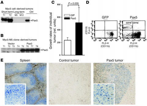
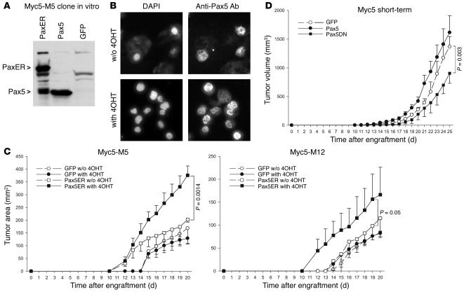
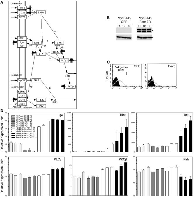
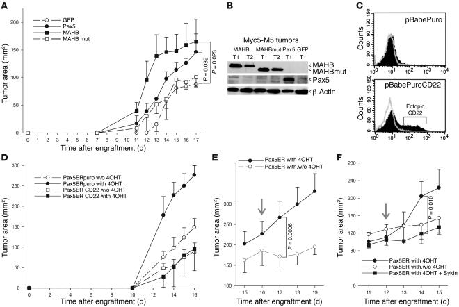
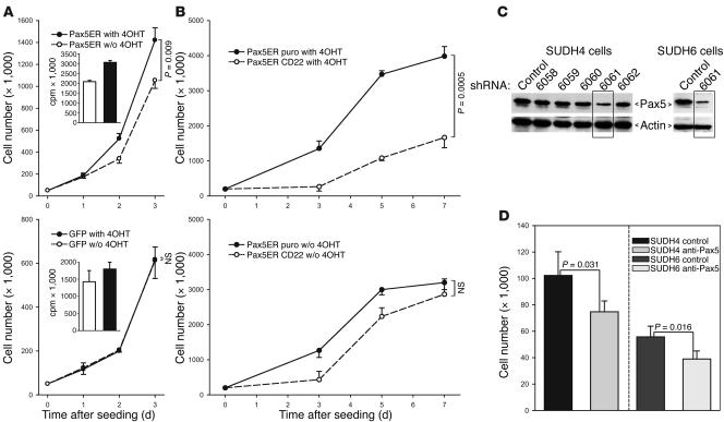
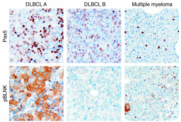
The presumed involvement of paired box gene 5 (PAX5) in B-lymphomagenesis is based largely on the discovery of Pax5-specific translocations and somatic hypermutations in non-Hodgkin lymphomas. Yet mechanistically, the contribution of Pax5 to neoplastic growth remains undeciphered. Here we used 2 Myc-induced mouse B lymphoma cell lines, Myc5-M5 and Myc5-M12, which spontaneously silence Pax5. Reconstitution of these cells with Pax5-tamoxifen receptor fusion protein (Pax5ER(TAM)) increased neoplastic growth in a hormone-dependent manner. Conversely, expression of dominant-negative Pax5 in murine lymphomas and Pax5 knockdown in human lymphomas negatively affected cell expansion. Expression profiling revealed that Pax5 was required to maintain mRNA levels of several crucial components of B cell receptor (BCR) signaling, including CD79a, a protein with the immunoreceptor tyrosine-based activation motif (ITAM). In contrast, expression of 2 known ITAM antagonists, CD22 and PIR-B, was suppressed. The key role of BCR/ITAM signaling in Pax5-dependent lymphomagenesis was corroborated in Syk, an ITAM-associated tyrosine kinase. Moreover, we observed consistent expression of phosphorylated BLNK, an activated BCR adaptor protein, in human B cell lymphomas. Thus, stimulation of neoplastic growth by Pax5 occurs through BCR and is sensitive to genetic and pharmacological inhibitors of this pathway.
Figures
Similar articles
-
CD19 is a major B cell receptor-independent activator of MYC-driven B-lymphomagenesis.J Clin Invest. 2012 Jun;122(6):2257-66. doi: 10.1172/JCI45851. Epub 2012 May 1. J Clin Invest. 2012. PMID: 22546857 Free PMC article.
-
B cell receptor-ERK1/2 signal cancels PAX5-dependent repression of BLIMP1 through PAX5 phosphorylation: a mechanism of antigen-triggering plasma cell differentiation.J Immunol. 2012 Jun 15;188(12):6127-34. doi: 10.4049/jimmunol.1103039. Epub 2012 May 16. J Immunol. 2012. PMID: 22593617
-
Differential PAX5 levels promote malignant B-cell infiltration, progression and drug resistance, and predict a poor prognosis in MCL patients independent of CCND1.Leukemia. 2016 Mar;30(3):580-93. doi: 10.1038/leu.2015.140. Epub 2015 May 15. Leukemia. 2016. PMID: 26073757 Free PMC article.
-
Diagnostic uses of Pax5 immunohistochemistry.Adv Anat Pathol. 2007 Sep;14(5):323-34. doi: 10.1097/PAP.0b013e3180ca8a49. Adv Anat Pathol. 2007. PMID: 17717432 Review.
-
The regulation of the B-cell gene expression programme by Pax5.Immunol Cell Biol. 2008 Jan;86(1):47-53. doi: 10.1038/sj.icb.7100134. Epub 2007 Nov 13. Immunol Cell Biol. 2008. PMID: 17998914 Review.
Cited by
-
A Myc-regulated transcriptional network controls B-cell fate in response to BCR triggering.BMC Genomics. 2009 Jul 17;10:323. doi: 10.1186/1471-2164-10-323. BMC Genomics. 2009. PMID: 19607732 Free PMC article.
-
Induction of Ca²+-driven apoptosis in chronic lymphocytic leukemia cells by peptide-mediated disruption of Bcl-2-IP3 receptor interaction.Blood. 2011 Mar 10;117(10):2924-34. doi: 10.1182/blood-2010-09-307405. Epub 2010 Dec 30. Blood. 2011. PMID: 21193695 Free PMC article.
-
Sellar B lymphoblastic lymphoma mimics pituitary apoplexy with newly discovered gene mutations in TP53 and PAX5: A case report.Front Oncol. 2023 Feb 7;13:1087232. doi: 10.3389/fonc.2023.1087232. eCollection 2023. Front Oncol. 2023. PMID: 36824134 Free PMC article.
-
2,3,7,8-Tetrachlorodibenzo-p-dioxin-mediated impairment of B cell differentiation involves dysregulation of paired box 5 (Pax5) isoform, Pax5a.J Pharmacol Exp Ther. 2008 Aug;326(2):463-74. doi: 10.1124/jpet.108.139857. Epub 2008 May 15. J Pharmacol Exp Ther. 2008. PMID: 18483191 Free PMC article.
-
PAX5 activates the transcription of the human telomerase reverse transcriptase gene in B cells.J Pathol. 2010 Jan;220(1):87-96. doi: 10.1002/path.2620. J Pathol. 2010. PMID: 19806612 Free PMC article.
References
-
- Shen-Ong G.L., Keath E.J., Piccoli S.P., Cole M.D. Novel myc oncogene RNA from abortive immunoglobulin-gene recombination in mouse plasmacytomas. Cell. 1982;31:443–452. - PubMed
-
- Adams J.M., et al. The c-myc oncogene driven by immunoglobulin enhancers induces lymphoid malignancy in transgenic mice. Nature. 1985;318:533–538. - PubMed
Publication types
MeSH terms
Substances
Grants and funding
LinkOut - more resources
Full Text Sources
Other Literature Sources
Medical
Miscellaneous
