. 2007 Sep;117(9):2713-22.
doi: 10.1172/JCI31457.

Mechanistic and prognostic significance of aberrant methylation in the molecular pathogenesis of human hepatocellular carcinoma

Affiliations

Mechanistic and prognostic significance of aberrant methylation in the molecular pathogenesis of human hepatocellular carcinoma

Diego F Calvisi et al. J Clin Invest. 2007 Sep.

Abstract

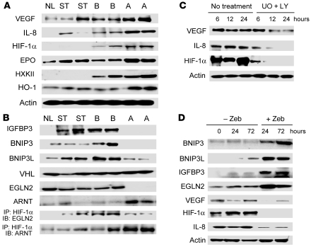
Hepatocellular carcinoma (HCC) is the fifth most common cancer worldwide, accounting for an estimated 600,000 deaths annually. Aberrant methylation, consisting of DNA hypomethylation and/or promoter gene CpG hypermethylation, is implicated in the development of a variety of solid tumors, including HCC. We analyzed the global levels of DNA methylation as well as the methylation status of 105 putative tumor suppressor genes and found that the extent of genome-wide hypomethylation and CpG hypermethylation correlates with biological features and clinical outcome of HCC patients. We identified activation of Ras and downstream Ras effectors (ERK, AKT, and RAL) due to epigenetic silencing of inhibitors of the Ras pathway in all HCC. Further, selective inactivation of SPRY1 and -2, DAB2, and SOCS4 and -5 genes and inhibitors of angiogenesis (BNIP3, BNIP3L, IGFBP3, and EGLN2) was associated with poor prognosis. Importantly, several epigenetically silenced putative tumor suppressor genes found in HCC were also inactivated in the nontumorous liver. Our results assign both therapeutic and chemopreventive significance to methylation patterns in human HCC and open the possibility of using molecular targets, including those identified in this study, to effectively inhibit HCC development and progression.


Figures

Figure 1
Figure 1. Analysis of DNA hypomethylation, degree of genomic instability, and CpG hypermethylation in normal livers (NL), nonneoplastic surrounding liver tissues (ST), and HCC with shorter (subclass A) and longer (subclass B) survival rate.
(A) As a measure of genome-wide hypomethylation, the incorporation of [3H]dCTP into DNA was assessed after treatment with HpaII endonuclease, cutting only nonmethylated sites. In nontumorous surrounding livers, global DNA hypomethylation was significantly higher than in normal livers. Genomic hypomethylation progressively increased from nonneoplastic surrounding livers to HCC. Liver tumors with poor prognosis displayed the highest levels of DNA hypomethylation. (B) Degree of genomic alterations as detected by RAPD analysis. The level of genomic alterations was significantly higher in subclass A than in subclass B HCC (P = 7.8 × 10⁻⁸). (C) Correlation between DNA hypomethylation and genomic alterations. r2 = 0.945; P < 0.001. (D) To determine CpG regional hypermethylation, DNA was pretreated with BssHII endonuclease. Since BssHII cuts only unmethylated sites, the lower [3H]dCTP incorporation indicates fewer unmethylated (cleaved) CpG sites and, therefore, a higher de novo methylation. CpG methylation levels were highest in HCC, particular in tumors of subclass A.
Figure 2
Figure 2. Summary of promoter hypermethylation frequency in subclass A and B HCC as detected by methylation-specific PCR.
Black and gray boxes indicate the presence or absence of promoter hypermethylation, respectively. Note the significantly higher number of genes affected by methylation in subclass A HCC. Only genes that were affected by promoter methylation in at least 1 sample are shown.
Figure 3
Figure 3. Methylation index in HCC from patients with shorter (subclass A) and longer (subclass B) survival.
Methylation index (or mean frequency of methylation) was defined as the ratio between the numbers of methylated genes to total number of examined genes in each sample. (A) Methylation index was highest in subclass A HCC. (B) Methylation index negatively correlated with the length of patient’s survival (r2 = 0.7387; P < 0.0001).
Figure 4
Figure 4. Activation of Ras and Ras downstream effectors in normal livers, nonneoplastic surrounding tissues, and HCC.
Activity of Ras (A), ERK (B), AKT (C), and Ral A (D) progressively increased from nontumorous surrounding livers to HCC, reaching the highest levels in subclass A HCC. Of note, the Ras downstream effector Ral A was active mostly in subclass A HCC. Proteins levels of total Ras (Pan Ras), ERK, AKT, and Ral A are shown to ensure equal protein loading. MBP, myelin basic protein; pGSK-3β, phospho–glycogen synthase kinase–3β.
Figure 5
Figure 5. Representative Western blot and immunoprecipitation analyses of Ras inhibitors and Ras downstream effectors in normal livers, nonneoplastic surrounding tissues, and HCC.
(A) Downregulation of EGFR inhibitors SOCS4 and SOCS5 resulted in increased EGFR levels, EGFR activation, and low EGFR/SOCS5 complexes in subclass A HCC. (B) Downregulation of SPRY1, -2, and -4 led to FGF receptor (FGFR) upregulation and increased FGF-mediated Ras signaling activation, as shown by a decrease in SPRY1/GRB2 and SPRY2/GRB2 complexes and increase in FRS2/GRB2 complexes in subclass A HCC. (C) In contrast, subclass B HCC showed elevated levels of both total (t) and membranous (m) SPRY1 and -2, leading to SPRY1 and -2 activation by tyrosine phosphorylation, increase in SPRY1/GRB2 and SPRY2/GRB2 complexes, and decrease in FRS2/GRB2 complexes (C). (D) DAB2 was downregulated in subclass A HCC, with consequent upregulation of c-Fos, Src, and ILK.
Figure 6
Figure 6. Representative Western blot and immuno­precipitation (IP) analyses of Ras proangiogenic targets (A) and inhibitors of Ras-driven angiogenesis (B).
Upregulation of HIF-1α, VEGF, and IL-8 (A) was associated with high HIF-1α/ARNT complexes, low levels of HIF-1α inhibitors (IGFBP3, BNIP3, BNIP3L, and EGLN2), and low EGLN2/HIF-1α complexes in subclass A HCC (B). (C) Suppression of Ras downstream effectors MEK (UO126 [UO]) and AKT (LY294002 [LY]) led to downregulation of HIF-1α, VEGF, and IL-8 in Focus hepatoma cell lines. (D) Reactivation of HIF-1α inhibitors and EGLN2 by Zebularine (Zeb) reduced HIF-1α, VEGF, and IL-8 expression in Focus hepatoma cell lines. Equivalent results were obtained in SNU-387 and 7703 hepatoma cell lines (data not shown).

References

    1. Bruix J., Boix L., Sala M., Llovet J.M. Focus on hepatocellular carcinoma. Cancer Cell. 2004;5:215–219. - PubMed
    1. Thorgeirsson S.S., Grisham J.W. Molecular pathogenesis of human hepatocellular carcinoma. Nat. Genet. 2002;31:339–346. - PubMed
    1. Baylin S.B. Mechanisms underlying epigenetically mediated gene silencing in cancer. Semin. Cancer Biol. 2002;12:331–337. - PubMed
    1. Jones P.A., Baylin S.B. The epigenomics of cancer. Cell. . 2007;128:683–692. - PMC - PubMed
    1. Wilson A.S., Power B.E., Molloy P.L. DNA hypomethylation and human diseases. Biochim. Biophys. Acta. 2007;1775:138–16. - PubMed

Publication types