

Intrathecal infusion of a Ca(2+)-permeable AMPA channel blocker slows loss of both motor neurons and of the astrocyte glutamate transporter, GLT-1 in a mutant SOD1 rat model of ALS
- PMID: 17719032
- PMCID: PMC2083564
- DOI: 10.1016/j.expneurol.2007.07.011
Intrathecal infusion of a Ca(2+)-permeable AMPA channel blocker slows loss of both motor neurons and of the astrocyte glutamate transporter, GLT-1 in a mutant SOD1 rat model of ALS
Abstract
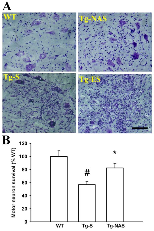
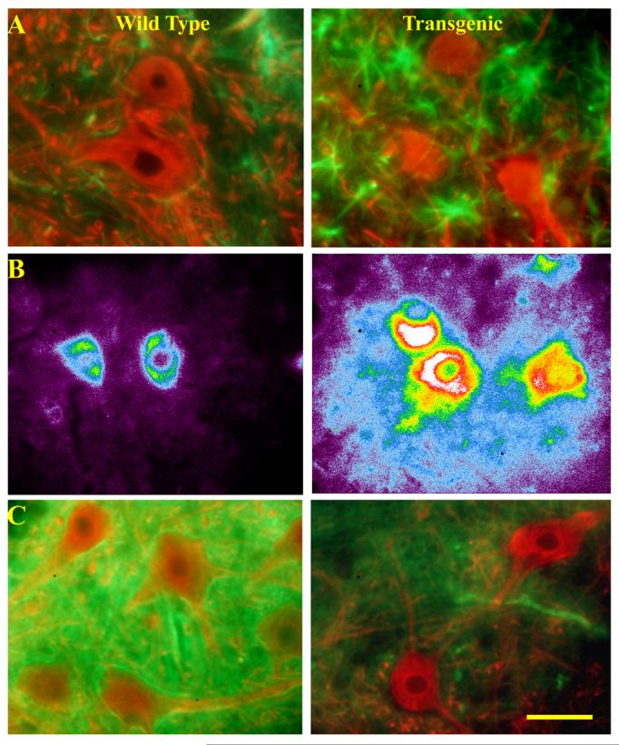
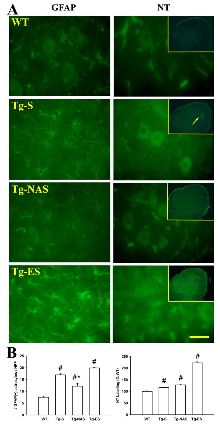
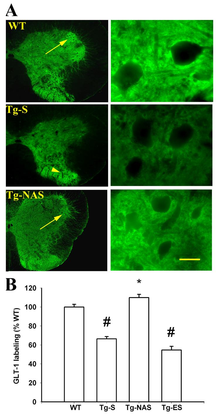
Elevated extracellular glutamate, resulting from a loss of astrocytic glutamate transport capacity, may contribute to excitotoxic motor neuron (MN) damage in ALS. Accounting for their high excitotoxic vulnerability, MNs possess large numbers of unusual Ca(2+)-permeable AMPA channels (Ca-AMPA channels), the activation of which triggers mitochondrial Ca(2+) overload and strong reactive oxygen species (ROS) generation. However, the causes of the astrocytic glutamate transport loss remain unexplained. To assess the role of Ca-AMPA channels on the evolution of pathology in vivo, we have examined effects of prolonged intrathecal infusion of the Ca-AMPA channel blocker, 1-naphthyl acetylspermine (NAS), in G93A transgenic rat models of ALS. In wild-type animals, immunoreactivity for the astrocytic glutamate transporter, GLT-1, was particularly strong around ventral horn MNs. However, a marked loss of ventral horn GLT-1 was observed, along with substantial MN damage, prior to onset of symptoms (90-100 days) in the G93A rats. Conversely, labeling with the oxidative marker, nitrotyrosine, was increased in the neuropil surrounding MNs in the transgenic animals. Compared to sham-treated G93A animals, 30-day NAS infusions (starting at 67+/-2 days of age) markedly diminished the loss of both MNs and of astrocytic GLT-1 labeling. These observations are compatible with the hypothesis that activation of Ca-AMPA channels on MNs contributes, likely in part through oxidative mechanisms, to loss of glutamate transporter in surrounding astrocytes.
Figures
Comment in
-
Crosstalk between astrocytes and motor neurons: what is the message?Exp Neurol. 2008 May;211(1):1-6. doi: 10.1016/j.expneurol.2008.01.008. Epub 2008 Jan 26. Exp Neurol. 2008. PMID: 18291372 Review.
Similar articles
-
Intrathecal infusion of BMAA induces selective motor neuron damage and astrogliosis in the ventral horn of the spinal cord.Exp Neurol. 2014 Nov;261:1-9. doi: 10.1016/j.expneurol.2014.06.003. Epub 2014 Jun 8. Exp Neurol. 2014. PMID: 24918341 Free PMC article.
-
Marked synergism between mutant SOD1 and glutamate transport inhibition in the induction of motor neuronal degeneration in spinal cord slice cultures.Brain Res. 2012 Apr 11;1448:153-62. doi: 10.1016/j.brainres.2012.02.005. Epub 2012 Feb 9. Brain Res. 2012. PMID: 22370146 Free PMC article.
-
Loss of metabotropic glutamate receptor-mediated regulation of glutamate transport in chemically activated astrocytes in a rat model of amyotrophic lateral sclerosis.J Neurochem. 2006 Feb;96(3):719-31. doi: 10.1111/j.1471-4159.2005.03577.x. Epub 2005 Dec 20. J Neurochem. 2006. PMID: 16371010
-
Transgenic SOD1 G93A mice develop reduced GLT-1 in spinal cord without alterations in cerebrospinal fluid glutamate levels.J Neurochem. 2001 Nov;79(4):737-46. doi: 10.1046/j.1471-4159.2001.00572.x. J Neurochem. 2001. PMID: 11723166
-
TNF-α triggers rapid membrane insertion of Ca(2+) permeable AMPA receptors into adult motor neurons and enhances their susceptibility to slow excitotoxic injury.Exp Neurol. 2012 Dec;238(2):93-102. doi: 10.1016/j.expneurol.2012.08.004. Epub 2012 Aug 19. Exp Neurol. 2012. PMID: 22921461 Free PMC article.
Cited by
-
Calcium dysregulation links ALS defective proteins and motor neuron selective vulnerability.Front Cell Neurosci. 2015 Jun 16;9:225. doi: 10.3389/fncel.2015.00225. eCollection 2015. Front Cell Neurosci. 2015. PMID: 26136661 Free PMC article. Review.
-
Cardiovascular Diseases, Medications, and ALS: A Population-Based Case-Control Study.Neuroepidemiology. 2022;56(6):423-432. doi: 10.1159/000526982. Epub 2022 Dec 8. Neuroepidemiology. 2022. PMID: 36481735 Free PMC article.
-
Intrathecal infusion of BMAA induces selective motor neuron damage and astrogliosis in the ventral horn of the spinal cord.Exp Neurol. 2014 Nov;261:1-9. doi: 10.1016/j.expneurol.2014.06.003. Epub 2014 Jun 8. Exp Neurol. 2014. PMID: 24918341 Free PMC article.
-
Ca permeable AMPA channels in diseases of the nervous system.Front Mol Neurosci. 2011 Nov 14;4:42. doi: 10.3389/fnmol.2011.00042. eCollection 2011. Front Mol Neurosci. 2011. PMID: 22102834 Free PMC article.
-
Intravenous injection of l-BMAA induces a rat model with comprehensive characteristics of amyotrophic lateral sclerosis/Parkinson-dementia complex.Toxicol Res (Camb). 2015 Nov 10;5(1):79-96. doi: 10.1039/c5tx00272a. eCollection 2016 Jan 1. Toxicol Res (Camb). 2015. PMID: 30090328 Free PMC article.
References
-
- Alexander GM, Deitch JS, Seeburger JL, Del Valle L, Heiman-Patterson TD. Elevated cortical extracellular fluid glutamate in transgenic mice expressing human mutant (G93A) Cu/Zn superoxide dismutase. J. Neurochem. 2000;74:1666–1673. - PubMed
-
- Beal MF, Ferrante RJ, Browne SE, Matthews RT, Kowall NW, Brown RH., Jr. Increased 3-nitrotyrosine in both sporadic and familial amyotrophic lateral sclerosis. Ann. Neurol. 1997;42:644–654. - PubMed
-
- Boillee S, Yamanaka K, Lobsiger CS, Copeland NG, Jenkins NA, Kassiotis G, Kollias G, Cleveland DW. Onset and progression in inherited ALS determined by motor neurons and microglia. Science. 2006;312:1389–1392. - PubMed
-
- Carriedo SG, Yin HZ, Lamberta R, Weiss JH. In vitro kainate injury to large, SMI-32(+) spinal neurons is Ca2+ dependent. Neuroreport. 1995;6:945–948. - PubMed
Publication types
MeSH terms
Substances
Grants and funding
LinkOut - more resources
Full Text Sources
Medical
Miscellaneous
