Comparative Study
. 2007 Aug 24;130(4):691-703.
doi: 10.1016/j.cell.2007.06.054.

Autocrine VEGF signaling is required for vascular homeostasis

Affiliations
Comparative Study

Autocrine VEGF signaling is required for vascular homeostasis

Sunyoung Lee et al. Cell. .

Abstract

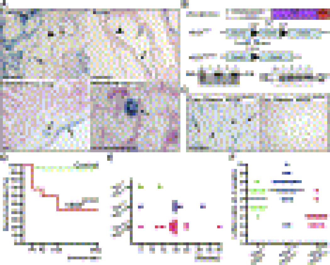
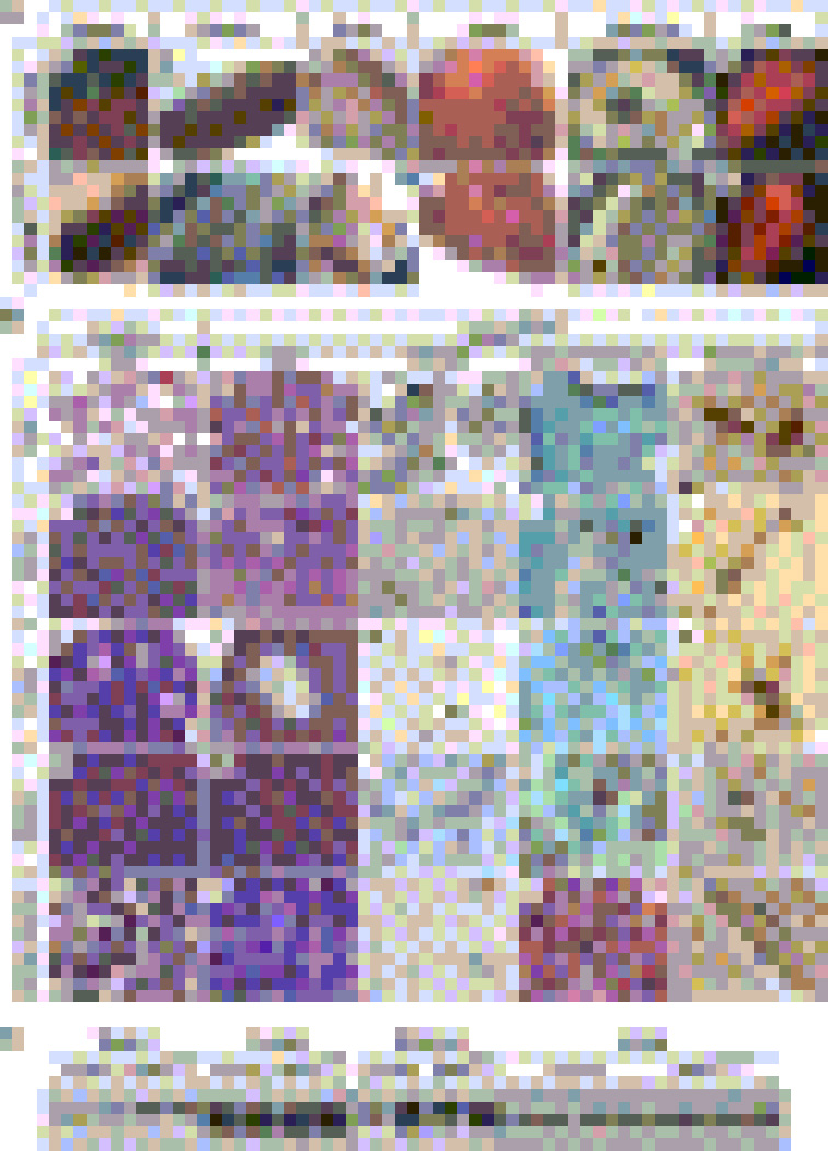
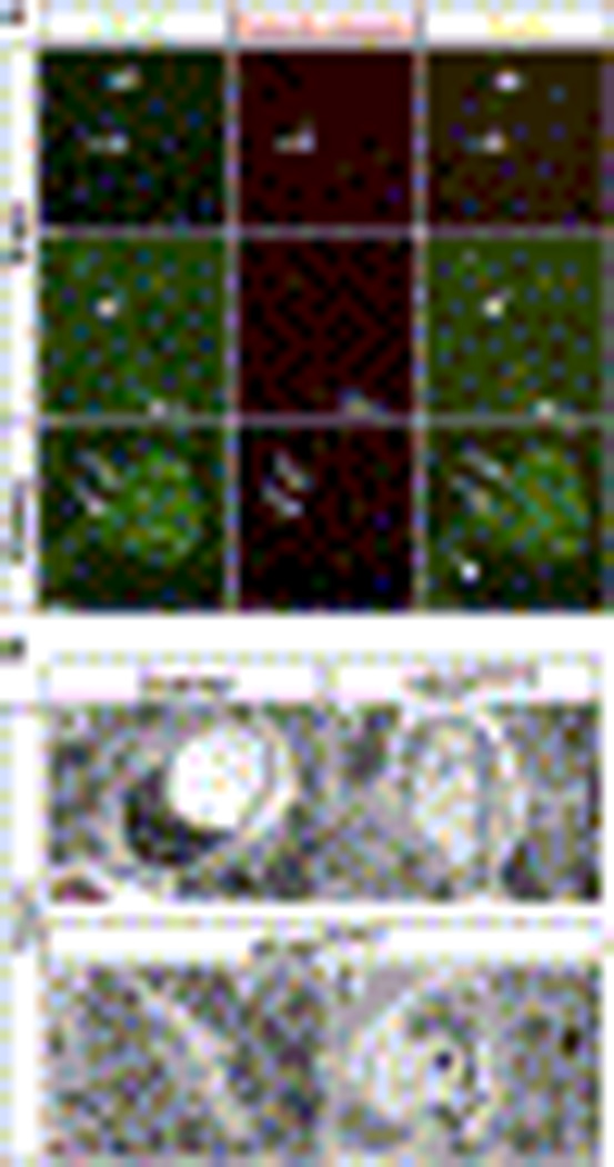
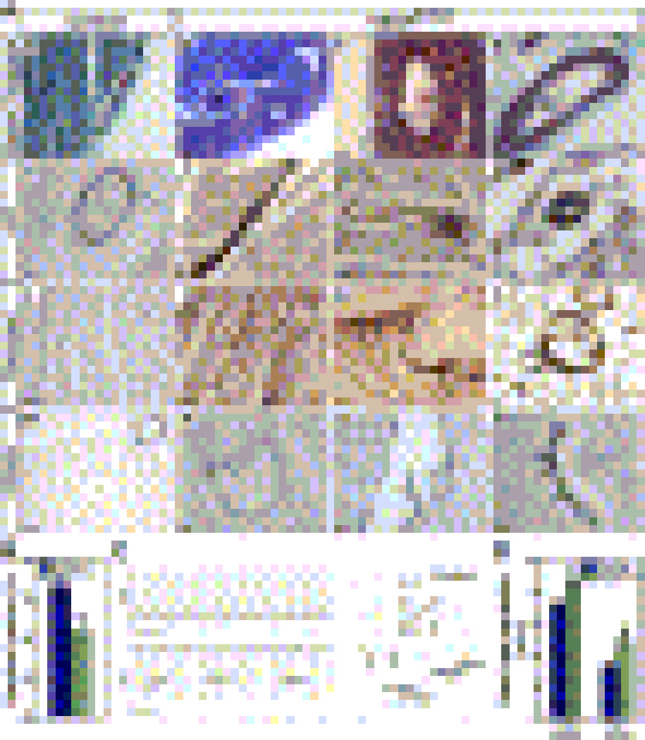
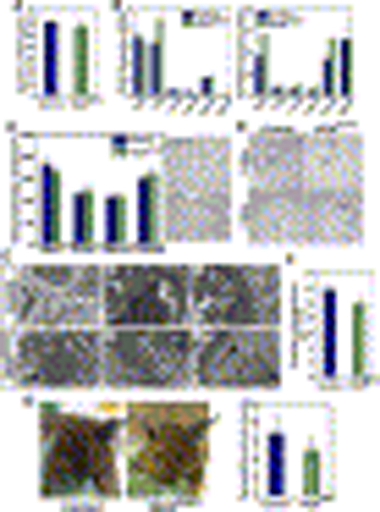
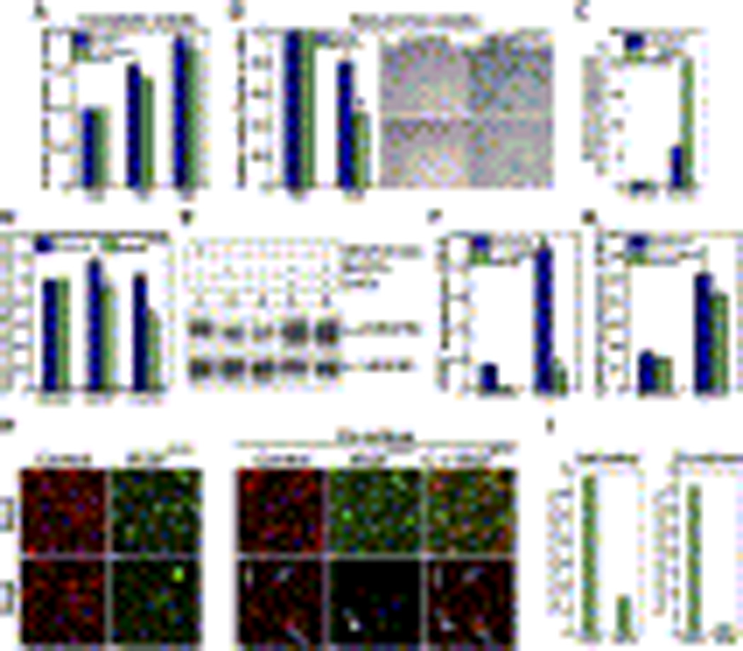
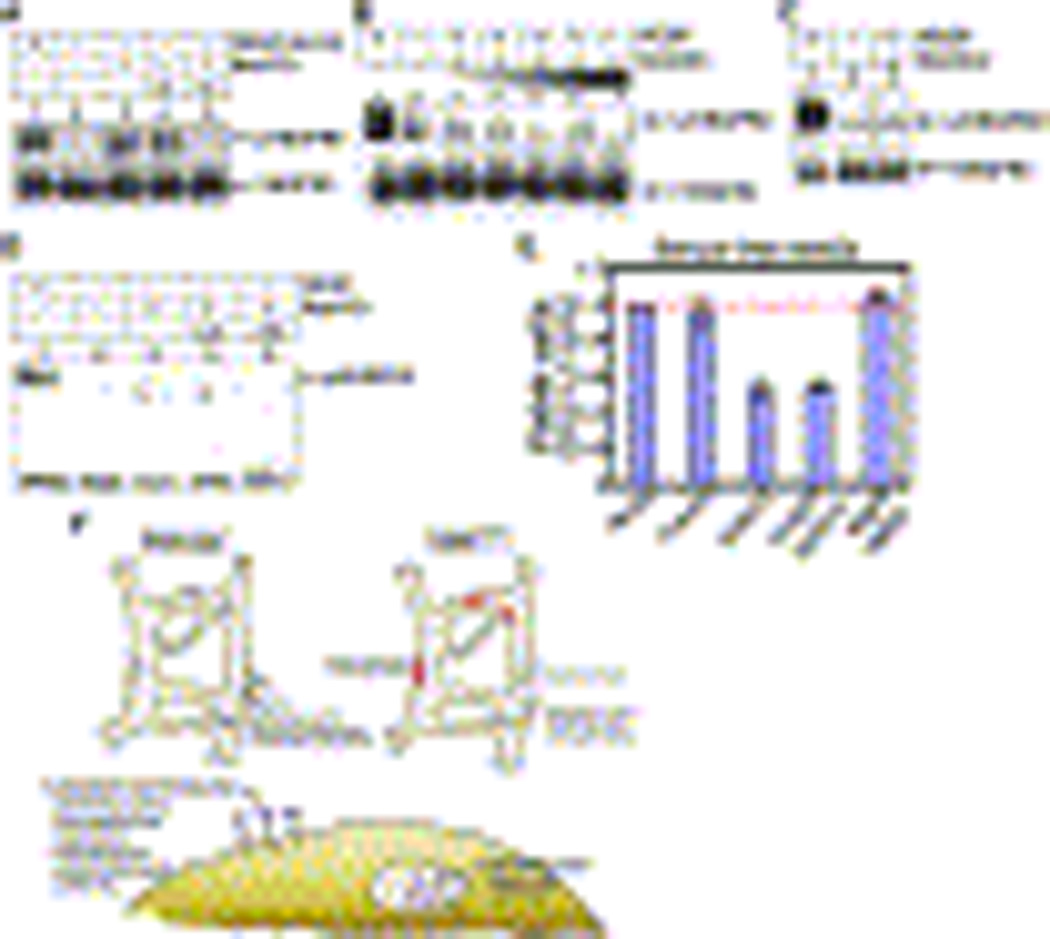
Vascular endothelial growth factor (VEGF) is essential for developmental and pathological angiogenesis. Here we show that in the absence of any pathological insult, autocrine VEGF is required for the homeostasis of blood vessels in the adult. Genetic deletion of vegf specifically in the endothelial lineage leads to progressive endothelial degeneration and sudden death in 55% of mutant mice by 25 weeks of age. The phenotype is manifested without detectable changes in the total levels of VEGF mRNA or protein, indicating that paracrine VEGF could not compensate for the absence of endothelial VEGF. Furthermore, wild-type, but not VEGF null, endothelial cells showed phosphorylation of VEGFR2 in the absence of exogenous VEGF. Activation of the receptor in wild-type cells was suppressed by small molecule antagonists but not by extracellular blockade of VEGF. These results reveal a cell-autonomous VEGF signaling pathway that holds significance for vascular homeostasis but is dispensable for the angiogenic cascade.


Figures

Figure 1
Figure 1. VEGF Expression by Endothelial Cells and Generation of VEGFECKO Mice
(A) Expression of LacZ reporter (blue) driven by the VEGF promoter in non-pathological adult tissues. Note promoter activity in endothelial cells (arrows). In larger vessels: positive (arrows) and negative (arrowhead) endothelial cells are observed. (B) Schematic diagram of the transgenic mice used to generate VEGFECKO mice (top). Genotyping of tail DNA. A band of 850 indicates the presence of the Cre transgene. Only animals from lanes 2 and 4 harbored the Cre-recombinase transgene (bottom, left). A 100-bp band indicates wild type (wt) allele and the 150-bp corresponds to the floxed VEGF allele (flox) (bottom, right). (C) Endothelial cells isolated from Cre+/Rosa+/VEGFlox/lox (VEGFECKO) and Cre-/Rosa+/VEGFlox/lox mice stained for β-gal. Arrows point to endothelial cells expressing Cre and arrowheads to cells with no Cre expression (approximately 95% of the isolated cells were positive). (D) Survival analysis. Mortality rate of newborn VEGFECKO at P0 was calculated by predicted mendelian distribution and expected litter size. Survival rate of VEGFECKO at six months was 34.7%. (E) Mortality of adult cohorts. (F) Litter size variability. Wt × Wt, 6 ± 1.33 (n = 10); Wt/KO × Wt/KO, 7.15 ± 1.76 (n = 20); KO/KO, 4.0 ± 1.30 (n = 11).
Figure 2
Figure 2. Systemic Vascular Pathologies VEGFECKO mice
(A) Macroscopic analyses. a–f, Control. g–l, VEGFECKO. VEGFECKO organs of older mice show hemorrhage (arrows in g, i and l), tortuous and dilated vessels (arrows in h), areas of suggestive collagen accumulation in the heart indicative of microinfarcts (arrows, j) and intestinal perforations (arrows, k) (also see histology in Fig. S1). (B) Histological sections of organs from younger mice. a–e, lung. Lung in VEGFECKO mice shows chronic inflammation (b), fibrosis (d) and excess of intravascular fibrin(ogen) deposits (e). PECAM staining reveals ruptured endothelial cells (arrowhead, c) and collapsed lumen (arrow, c). f–j, uterus. k–o, ovary. VEGFECKO ovary shows significantly enlarged vessels surrounding mature follicles (l) compared to wild type ovary (k). p–t, spleen. Asterisk indicates fibrosis. u–y, bone marrow. Fibrin(ogen) staining reveals clotting from VEGFECKO organs (arrows, e, o, t and y). Hemosiderin deposits were indicated by arrows in h, i, m, n, r, s, w and x. Bar, 100µm. (C) VEGFR2 protein levels from VEGFECKO and control at 25 weeks were determined by immunoblots (same amount of protein was loaded per well). Slight differences in the uterus correlate with estrous cycle.
Figure 3
Figure 3. Evidence of Endothelial Cell Apoptosis in VEGFECKO Mice
(A) Endothelial expression of cleaved caspase-3 in VEGFECKO mice. Representative sections double-stained for PECAM (green) and cleaved caspase-3 (red) are shown. Arrows point to vessels with active caspase-3. Arrowheads indicate caspase-3 negative vessels. Bar, 100µm. (B) Electron microscopic evidence of endothelial cell apoptosis in heart sections. Endothelial cells of VEGFECKO heart (b–d) shows morphological characteristic of apoptotic cells, including cytoplasmic swelling (b–c, arrow), nuclear condensation (d, arrow) and the exposure of cytosolic components (d, asterisk). Also note mitochondria swelling in d (arrowhead). Bar, 3.5µm
Figure 4
Figure 4. Intravascular Thrombosis in VEGFECKO Mice Affect Cardiac Physiology
(A) a–d, Frequent intravascular thrombi (b, asterisks), detachment of endothelial lining (c, arrow) and platelet adhesion (d, arrows) in VEGFECKO hearts. (a–d, Gomori trichrome staining). e–h, von-Willebrand Factor (vWF) staining shows accumulation in the vascular wall of mutant mice (arrows). i–l. Fibrinogen staining was also increased (arrows). m–p, PECAM staining shows detachment of endothelial cells (n, o, arrows) and collapse of vascular wall (p, arrows). Bars: a–c, f, g, k, m, p= 35µm; d, e, h, l, n, o, p= 10µm. (B) VEGFECKO mice (green) have reduced ejection fraction compared to control littermates (blue) (means ± SD., n=11 ea, P<0.001). (C) ECG traces obtained from wild type (top) and VEGFECKO (bottom) mice. The left-right arrows in VEGFECKO (bottom) mice indicate the variability in heart rate and amplitude. Time bar = 0.5 sec. The right panels show expanded ECG traces with the defections annotated. Time bar = 0.5 sec. (D) Increased heart rate variability from wild type (black) to VEGFECKO (green) mice during both diurnal cycles (mean ± SD., n=3 ea, P<0.001).
Figure 5
Figure 5. VEGF Levels, Vessel Density, Endothelial Fenestrations and Vessel Permeability in VEGFECKO and Control Mice
(A) Serum VEGF levels from wild type (blue) and VEGFECKO (green) mice were determined by ELISA. (B) Organs from wild type (blue) and VEGFECKO (green) mice were analyzed for VEGF (left) and GAPDH (right) transcripts by real-time RT-PCR. Relative RNA units (RRU) were normalized to β-actin levels and calculated from standard curves. (C) Assessment of vascular density. Data shown are means (n=4) ± S.D (left). Representative PECAM-stained sections of uterus is shown on the right. (D) Induction of angiogenesis in wild type and VEGFECKO using Matrigel plugs containing VEGF. Arrows indicate neovessels invading the Matrigel plug. (E) Ultrastructural analyses of glomerular capillaries. Arrows show fenestrations in control and VEGFECKO mice. Note the swelling of VEGFECKO endothelium (asterisk in d and closed triangle in f), yet these cells still retain fenestrations (arrows). (F) Quantification of fenestrations in control and VEGFECKO mice. Glomerular fenestrations from wild type (blue) and VEGFECKO (green) mice were determined using ultrastructural information obtained from 4 mice. Data shown are means ± SD. Evaluation of P value indicates that the difference is statistically insignificant. (G) Vascular permeability responses. (left) Photographs of control and VEGFECKO mice injected with Evans Blue followed by application of vehicle or mustard oil (arrow) in the ear. (right) Quantification of extravasated Evans Blue dye from wild type (blue) and VEGFECKO (green) mice. Data are presented as ratio to control. P value was not significant.
Figure 6
Figure 6. Endothelial VEGF Mediates Survival in a Cell-Autonomous Manner
(A–B) Endothelial cells from control (blue) and VEGFECKO (green) mice were cultured in the presence (A) or absence (B) of serum and viable cells were counted at indicated times. Endothelial cells were stained for x-gal and nuclear fast red (right panels, B). Note the difference in cell number between control and VEGFECKO cells. (C) Endothelial cells from control (blue) and VEGFECKO (green) mice were cultured in the presence of CoCl2, active caspases were fluorescence-labeled and quantified by fluorometer. (D) Endothelial cells from control (blue) and VEGFECKO (green) mice were cultured in the presence of recombinant hVEGF165 or CoCl2 and viable cells assessed after 48hs. (E) HUVECs were cultured under hypoxia for 24 h and analyzed for VEGFR2 phosphorylation (pVEGFR2, top) and VEGFR2 total levels (bottom). Lanes: 1, VEGF (100 ng/ml) for 5 min; 2–3, normoxia in the presence (2) and absence (3) of sodium orthovanadate (Na3VO4) for 24 h; 4–5, CoCl2 (hypoxia mimetic) in the presence of Na3VO4 for 24 h. (F–G) Endothelial cells from control (blue) and VEGFECKO (green) mice were cultured in the presence of CoCl2 (100 µM) for 24 h and transcript levels of VEGF (F) and GAPDH (G) were determined by real-time RT-PCR. Relative RNA units (RRU) were normalized to β-actin. Data shown are means ± SD. (H) Endothelial cells differentially labeled (Cy3 for wild type and BODIFY green for VEGFECKO) were cultured alone (left) or combined (right). Photographs were taken after 2 days and 5 days. Arrows indicate control cells (red). Arrowhead points to VEGFECKO cells (green). (I) Quantification of cell survival following co-culture. Fluorescence labeled cells were counted at indicated times. Data are presented as percentage to control.
Figure 7
Figure 7. Autocrine VEGF Signaling in Endothelial Cells
(A) HUVECs were cultured under normoxia for 24 h and in the presence of the indicated compounds and analyzed for VEGFR2 phosphorylation (top) and VEGFR2 total levels (bottom). Lanes: 1, 100 ng/ml VEGF for 5 min; 2, media; 3, Na3VO4; 4–5, Avastin (10 µg/ml, 4) or SU4312 (0.4 µM, 5) in the presence of Na3VO4. (B) HUVECs were exposed to VEGF (100 ng/ml, pre-incubated with Avastin at 37°C for 2 h) for 5 min. Phosphorylation of VEGFR2 (top) and VEGFR2 (bottom) are shown. Lanes: 1, VEGF; 2, media; 3–6, VEGF pre-incubated with 1 ug/ml Avastin (3), 5 ug/ml Avastin (4), 10 ug/ml Avastin (5) and 100 ug/ml Avastin (6); 7, Avastin (1 ug/ml). (C) HUVECs incubated with VEGF (100 ng/ml, lane 1) or vehicle (lane 2). In lane 3, HUVECs were pre-incubated with SU4312 at 37°C for 2 h and then exposed to VEGF (100 ng/mlPhosphorylation of VEGFR2 (top) and total levels of VEGFR2 (bottom) are shown. (D) Endothelial cells from VEGFECKO were cultured under normoxia for 24 h and subjected to the indicated treatments. Lysates were analyzed for VEGFR2 phosphorylation. Lanes: 1, VEGF (100 ng/ml) for 5 min; 2, media; 3, Na3VO4; 4–5, Avastin (10 µg/ml, 4) or SU4312 (0.4 µM, 5) in the presence of Na3VO4. (E) HUVECs were cultured in the absence of serum with indicated VEGF or VEGF signaling blockers. (F) Schematic representation of VEGF function in endothelial homeostasis. Stress induced by radiation, reactive-oxygen species and hypoxia triggers activation of VEGFR2 by both autocrine and intracrine VEGF sources supporting endothelial survival. In the absence of intracrine VEGF, some endothelial cells undergo apoptosis resulting in hemorrhage (smaller vessels), and / or exposure of the underlying basement membrane with subsequent development of thrombi (larger vessels). In the bottom, the scheme shows an endothelial cell in which paracrine and intra/autocrine activation is taking place.

Comment in

Similar articles

Cited by

References

    1. Alva JA, Zovein AC, Monvoisin A, Murphy T, Salazar A, Harvey NL, Carmeliet P, Iruela-Arispe ML. VE-Cadherin-Cre-recombinase transgenic mouse: a tool for lineage analysis and gene deletion in endothelial cells. Dev Dyn. 2006;235:759–767. - PubMed
    1. Bates D, Taylor GI, Minichiello J, Farlie P, Cichowitz A, Watson N, Klagsbrun M, Mamluk R, Newgreen DF. Neurovascular congruence results from a shared patterning mechanism that utilizes Semaphorin3A and Neuropilin-1. Dev Biol. 2003;255:77–98. - PubMed
    1. Blann AD. Assessment of endothelial dysfunction: focus on atherothrombotic disease. Pathophysiol Haemost Thromb. 2003a;33:256–261. - PubMed
    1. Blann AD. How a damaged blood vessel wall contibutes to thrombosis and hypertenasion. Pathophysiol Haemost Thromb. 2003b;33:445–448. - PubMed
    1. Bokel C, Schwabedissen A, Entchev E, Renaud O, Gonzalez-Gaitan M. Sara endosomes and the maintenance of Dpp signaling levels across mitosis. Science. 2006;314:1135–1139. - PubMed

Publication types

MeSH terms