Review
. 2007 Oct;43(4):388-403.
doi: 10.1016/j.yjmcc.2007.07.045. Epub 2007 Jul 21.

Factors controlling cardiac myosin-isoform shift during hypertrophy and heart failure

Affiliations
Review

Factors controlling cardiac myosin-isoform shift during hypertrophy and heart failure

Mahesh P Gupta. J Mol Cell Cardiol. 2007 Oct.

Abstract

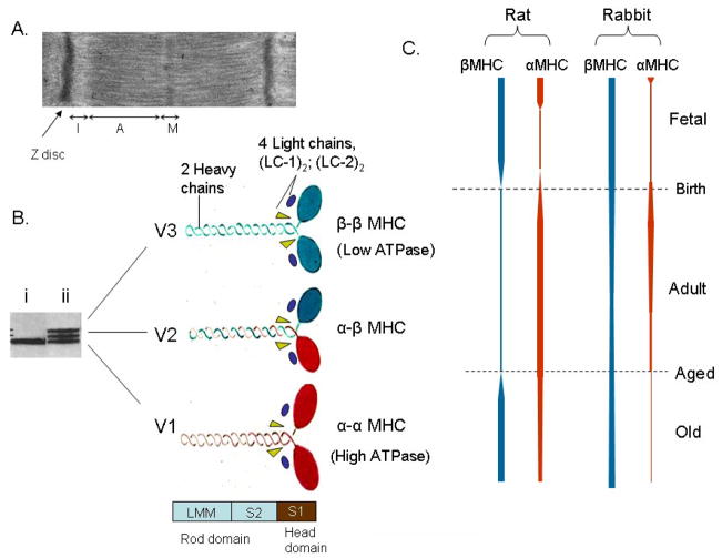
Myosin is a molecular motor, which interacts with actin to convert the energy from ATP hydrolysis into mechanical work. In cardiac myocytes, two myosin isoforms are expressed and their relative distribution changes in different developmental and pathophysiologic conditions of the heart. It has been realized for a long time that a shift in myosin isoforms plays a major role in regulating myocardial contractile activity. With the recent evidence implicating that alteration in myosin isoform ratio may be eventually beneficial for the treatment of a stressed heart, a new interest has developed to find out ways of controlling the myosin isoform shift. This article reviews the published data describing the role of myosin isoforms in the heart and highlighting the importance of various factors shown to influence myosin isofrom shift during physiology and disease states of the heart.


Figures

Figure 1
Figure 1
Structural organization and developmental regulation of MHC isoforms. (A) Transmission electron micrograph of rat heart muscle showing different regions of a sarcomere. MHC isoforms are located in the “A” band of the sarcomere. (B) A native gel showing separation of three ventricular myosin isoforms of an old aged (ii) and young adult (i) rat heart [17]. The composition of three (V1, V2 & V3) isofroms made up of 2 heavy chain subunits and 4 light chains is shown. Proteolytic fragment S1, corresponding to myosin head domain and two other proteolytic fragments, S2 and LMM (light meromyosins) corresponding to alpha-helical rod domain are shown. (C) Schematic representation of the developmental regulation of rat and rabbit heart myosin isoforms during fetal, adult and old age of the animal [15, 16, 154].
Figure 2
Figure 2
Models to explain HDAC-mediated regulation of MHC gene expression during hypertrophy. (A) Induction of βMHC gene expression by blocking the activity of class-II HDACs (HDAC 4/5). Class-II HDACs binds to SRF and MEF2 leading to repression of their gene transcription activity. Hypertrophy stimuli activate the activity of a kinase (Ca/calmodulin dependent kinase and/or HDAC kinase), which phosphorylates two serine residues conserved in class-II HDACs [128]. Upon phosphorylation, class-II HDACs are exported out from nucleus to cytoplasm, resulting in the de-repression of SRF and MEF2 transcription activity and induction of the βMHC expression from SRF and MEF2 binding sites [127, 128]. In a parallel pathway, activation of the PK-c signaling phosphorylates TEF1 leading to activation of its gene activation potential. TEF1 physically binds and functionally interacts with SRF and MEF2. It is therefore likely that a combined effect of these three regulators of gene expression controls the βMHC gene induction in response to hypertrophic stimuli. (B) Activation of αMHC gene expression by trichostatin A (TSA). The effect of TSA on αMHC induction is mediated by inhibition of the class-I HDACs and it occurs in euthyroid and not in hypothyroid animals (see text). In the absence of ligand (T3), thyroid receptor (TR) interacts with a multiprotein co-repressor (CoR) complex that contains class-I HDACs. This complex represses the gene transcription. In the presence of ligand (T3), the TR undergoes a conformational change, which results in replacement of the co-repressor complex with an activator complex (CoA) that contains HATs (p300/PCAF). Inhibition of HDACs by TSA enhances the activity of this activator complex with TR, resulting in the induction of the α-MHC gene expression. The activity of αMHC gene promoter is also controlled by change in activity of PARP1, which is activated by elevated levels of Ca2+ and increased activity of MEK/ERK1/2 signaling. PARP1 forms a repressor complex with class-I HDACs. Thus, TSA can also activate the αMHC gene expression by blocking the repressor activity of PARP1/class-I HDACs complex. In this context, another MHC gene regulator, TEF1, which physically binds to PARP1 can also modulate the PARP activity. TEF1 is phosphorylated by PK-A, and that stimulates its gene transcription activity [80]. Thus, we propose that the activation of PARP1 by TSA, and the activation of TEF1 by PK-A-signaling may have a co-operative effect for the activation of the αMHC gene expression (for details see text).
Figure 3
Figure 3
Increased activity of PARP1 in physiologic and pathologic hypertrophy. (A) Heart nuclear extract of mice subjected to swimming or aortic banding was analyzed by western analysis using anti-poly (ADP) ribose antibody. (B) Linear-regression analysis of PARP activity with the degree of cardiac hypertrophy. These studies are previously published in ref [107].

Similar articles

Cited by

References

    1. Spann J. Functional changes o in pathologic hypertrophy. In: Zak R, editor. Growth of the Heart in Health and Disease. New York: Raveen Press; 1984. pp. 421–466.
    1. Hajjar RJ, Gwathmey JK. Cross-bridge dynamics in human ventricular myocardium. Regulation of contractility in the failing heart. Circulation. 1992;86:1819–26. - PubMed
    1. Mann DL, Urabe Y, Kent RL, Vinciguerra S, Cooper G. Cellular versus myocardial basis for the contractile dysfunction of hypertrophied myocardium. Circ Res. 1991;68:402–15. - PubMed
    1. Shroff SG, Motz W. Left ventricular systolic resistance in rats with hypertension and hypertrophy. Am J Physiol. 1989;257:H386–94. - PubMed
    1. Anderson RL, Kawas RF, Pokrovskii MV, Godinez G, Lee JK, Mak J, et al. The cardaic myosin activator CK-1316719 increases myofibril ATPase activity and myocyte contractility in a rat model of heart failure. Circulation. 2006;114(suppl II):1440.

Publication types

MeSH terms