Increased apoptosis and myocyte enlargement with decreased cardiac mass; distinctive features of the aging male, but not female, monkey heart
- PMID: 17720187
- PMCID: PMC2701621
- DOI: 10.1016/j.yjmcc.2007.07.048
Increased apoptosis and myocyte enlargement with decreased cardiac mass; distinctive features of the aging male, but not female, monkey heart
Abstract
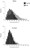
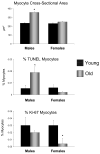
We studied gender-specific changes in aging cardiomyopathy in a primate model, Macaca fascicularis, free of the major human diseases, complicating the interpretation of data specific to aging in humans. Left ventricular (LV) weight/body weight decreased, p<0.05, in old males but did not change in old females. However, despite the decrease in LV weight, mean myocyte cross-sectional area in the old males increased by 51%. This increase in myocyte size was not uniform in old males, i.e., it was manifest in only 20-30% of all the myocytes from old males. In old males there was a 4-fold increase in frequency of myocyte apoptosis without any increase in proliferation-capable myocytes assessed by Ki-67 expression. Apoptosis was unchanged in old female monkey hearts, whereas the frequency of myocytes expressing Ki-67 declined 90%. These results, opposite to findings from rodent studies, indicate distinct differences in which male and female monkeys maintain functional heart mass during aging. The old male hearts demonstrated increased apoptosis, which more than offset the myocyte hypertrophy. Interestingly, the hypertrophy was not uniform and there was no significant increase in myocyte proliferation.
Figures


Similar articles
-
Aging, cardiac hypertrophy and ischemic cardiomyopathy do not affect the proportion of mononucleated and multinucleated myocytes in the human heart.J Mol Cell Cardiol. 1996 Jul;28(7):1463-77. doi: 10.1006/jmcc.1996.0137. J Mol Cell Cardiol. 1996. PMID: 8841934
-
Novel mechanisms for caspase inhibition protecting cardiac function with chronic pressure overload.Basic Res Cardiol. 2013 Jan;108(1):324. doi: 10.1007/s00395-012-0324-y. Epub 2013 Jan 1. Basic Res Cardiol. 2013. PMID: 23277091 Free PMC article.
-
Growth hormone increases the proliferation of existing cardiac myocytes and the total number of cardiac myocytes in the rat heart.Cardiovasc Res. 2007 Dec 1;76(3):400-8. doi: 10.1016/j.cardiores.2007.06.026. Epub 2007 Jul 4. Cardiovasc Res. 2007. PMID: 17673190
-
Evaluation of myocyte proliferation in alcoholic cardiomyopathy: telomerase enzyme activity (TERT) compared with Ki-67 expression.Alcohol Alcohol. 2011 Sep-Oct;46(5):534-41. doi: 10.1093/alcalc/agr071. Epub 2011 Jul 5. Alcohol Alcohol. 2011. PMID: 21733836
-
Apoptosis in heart failure.Prog Cardiovasc Dis. 1998 May-Jun;40(6):549-62. doi: 10.1016/s0033-0620(98)80003-0. Prog Cardiovasc Dis. 1998. PMID: 9647609 Review.
Cited by
-
Myocardial extracellular volume fraction from T1 measurements in healthy volunteers and mice: relationship to aging and cardiac dimensions.JACC Cardiovasc Imaging. 2013 Jun;6(6):672-83. doi: 10.1016/j.jcmg.2012.09.020. Epub 2013 May 1. JACC Cardiovasc Imaging. 2013. PMID: 23643283 Free PMC article.
-
Factors Associated With the Improvement of Left Ventricular Systolic Function by Continuous Positive Airway Pressure Therapy in Patients With Heart Failure With Reduced Ejection Fraction and Obstructive Sleep Apnea.Front Neurol. 2022 Mar 10;13:781054. doi: 10.3389/fneur.2022.781054. eCollection 2022. Front Neurol. 2022. PMID: 35359656 Free PMC article.
-
Cardiac proteasome activity in muscle ring finger-1 null mice at rest and following synthetic glucocorticoid treatment.Am J Physiol Endocrinol Metab. 2011 Nov;301(5):E967-77. doi: 10.1152/ajpendo.00165.2011. Epub 2011 Aug 9. Am J Physiol Endocrinol Metab. 2011. PMID: 21828340 Free PMC article.
-
Unlocking the dark matter: noncoding RNAs and RNA modifications in cardiac aging.Am J Physiol Heart Circ Physiol. 2024 Mar 1;326(3):H832-H844. doi: 10.1152/ajpheart.00532.2023. Epub 2024 Feb 2. Am J Physiol Heart Circ Physiol. 2024. PMID: 38305752 Free PMC article. Review.
-
Role of Beta-adrenergic Receptors and Sirtuin Signaling in the Heart During Aging, Heart Failure, and Adaptation to Stress.Cell Mol Neurobiol. 2018 Jan;38(1):109-120. doi: 10.1007/s10571-017-0557-2. Epub 2017 Oct 24. Cell Mol Neurobiol. 2018. PMID: 29063982 Free PMC article. Review.
References
-
- Lakatta EG. Cardiovascular ageing in health sets the stage for cardiovascular disease. Heart Lung Circ. 2002;11:76–91. - PubMed
-
- Lakatta EG. Arterial and cardiac aging: major shareholders in cardiovascular disease enterprises: Part III: cellular and molecular clues to heart and arterial aging. Circulation. 2003;107:490–7. - PubMed
-
- Lakatta EG, Levy D. Arterial and cardiac aging: major shareholders in cardiovascular disease enterprises: Part II: the aging heart in health: links to heart disease. Circulation. 2003;107:346–54. - PubMed
-
- Lakatta EG, Levy D. Arterial and Cardiac Aging: Major Shareholders in Cardiovascular Disease Enterprises: Part I: Aging Arteries: A “Set Up” for Vascular Disease. Circulation. 2003;107:139–146. - PubMed
-
- Takagi G, Asai K, Vatner SF, Kudej RK, Rossi F, Peppas A, et al. Gender differences on the effects of aging on cardiac and peripheral adrenergic stimulation in old conscious monkeys. Am J Physiol Heart Circ Physiol. 2003;285:H527–34. - PubMed
Publication types
MeSH terms
Substances
Grants and funding
- R37 HL033107/HL/NHLBI NIH HHS/United States
- HL069752/HL/NHLBI NIH HHS/United States
- AG014121/AG/NIA NIH HHS/United States
- P01 AG027211/AG/NIA NIH HHS/United States
- T32 HL069752/HL/NHLBI NIH HHS/United States
- R01 HL033107/HL/NHLBI NIH HHS/United States
- HL069020/HL/NHLBI NIH HHS/United States
- AG023567/AG/NIA NIH HHS/United States
- P01 HL069020/HL/NHLBI NIH HHS/United States
- P01 HL059139/HL/NHLBI NIH HHS/United States
- R01 AG023137/AG/NIA NIH HHS/United States
- HL059139/HL/NHLBI NIH HHS/United States
- HL033107/HL/NHLBI NIH HHS/United States
- R01 AG014121/AG/NIA NIH HHS/United States
- AG023137/AG/NIA NIH HHS/United States
- AG028854/AG/NIA NIH HHS/United States
- R01 AG028854/AG/NIA NIH HHS/United States
LinkOut - more resources
Full Text Sources
Medical

