

The antivascular action of physiotherapy ultrasound on a murine tumor: role of a microbubble contrast agent
- PMID: 17720299
- PMCID: PMC2423191
- DOI: 10.1016/j.ultrasmedbio.2007.06.013
The antivascular action of physiotherapy ultrasound on a murine tumor: role of a microbubble contrast agent
Abstract
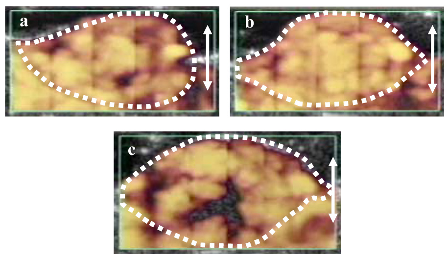
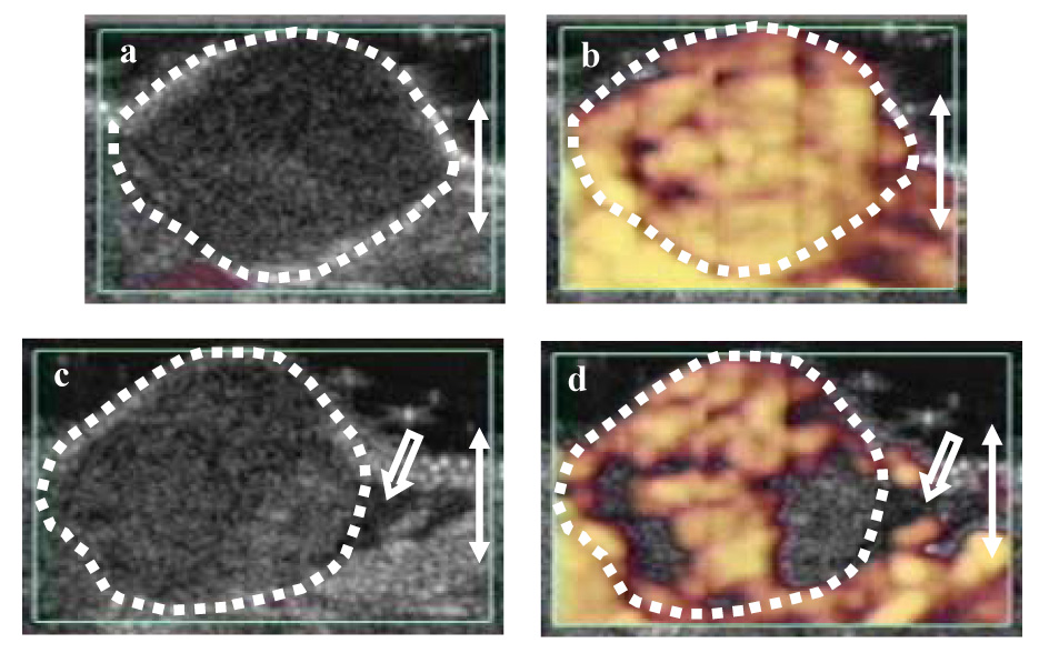
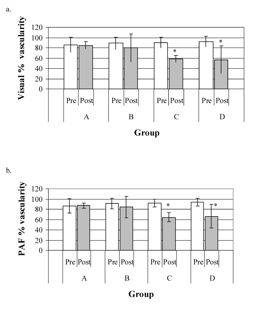
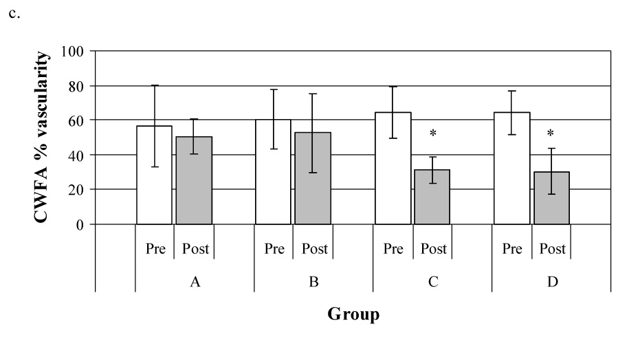
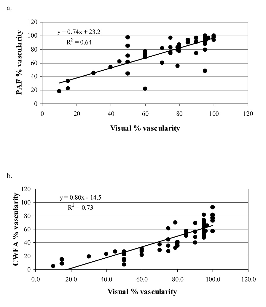
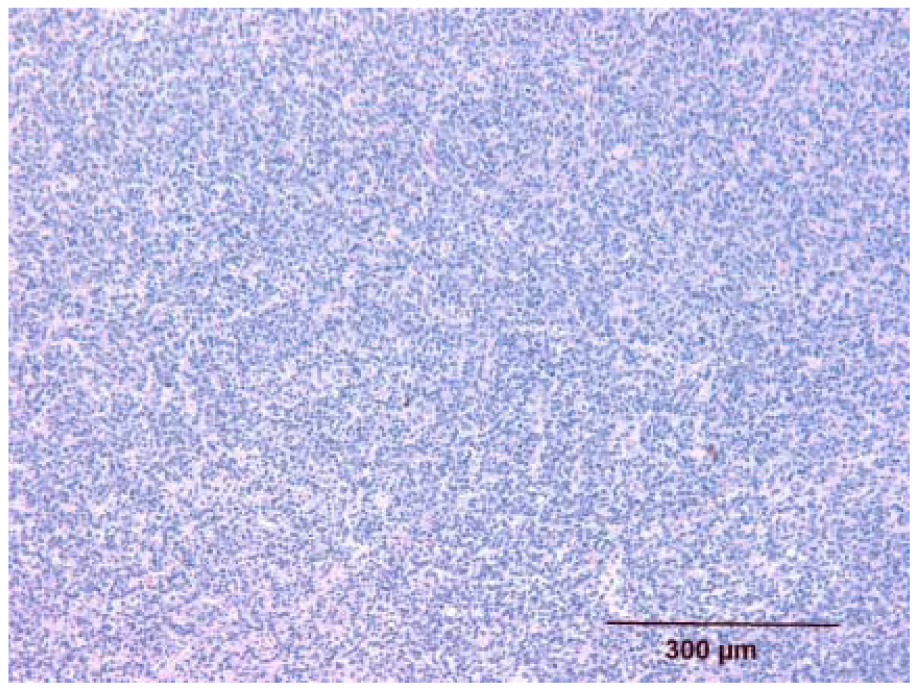
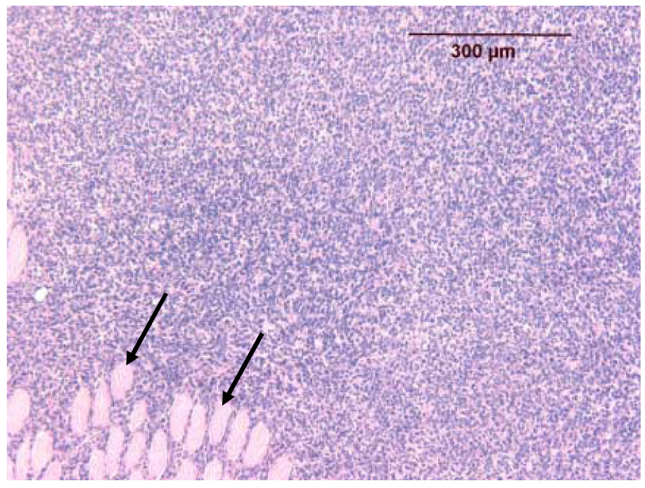
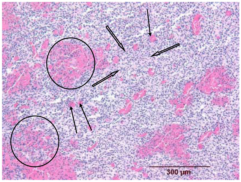
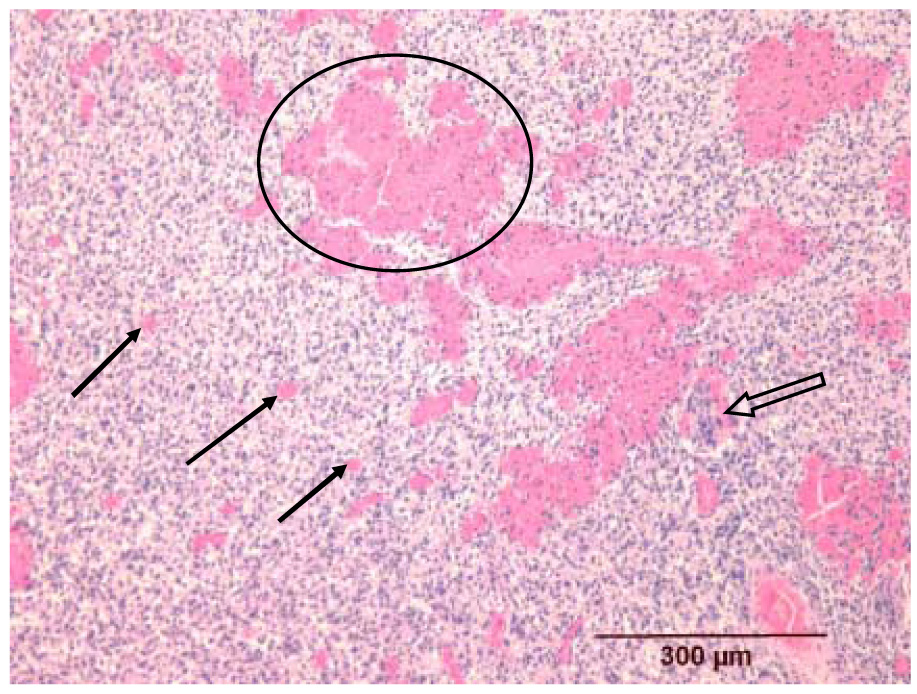
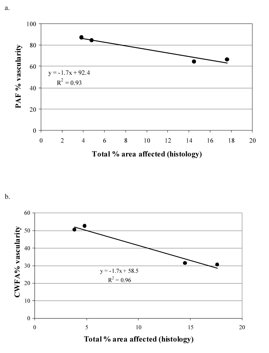
This study investigated whether a microbubble-containing ultrasound contrast agent had a role in the antivascular action of physiotherapy ultrasound on tumor neovasculature. Ultrasound images (B-mode and contrast-enhanced power Doppler [0.02 mL Definity]) were made of 22 murine melanomas (K1735(22)). The tumor was insonated (I(SATA) = 1.7 W cm(-2), 1 MHz, continuous output) for 3 min and the power Doppler observations of the pre- and postinsonation tumor vascularities were analyzed. Significant reductions (p = 0.005 for analyses of color-weighted fractional area) in vascularity occurred when a contrast-enhanced power Doppler study occurred before insonation. Vascularity was unchanged in tumors without a pretherapy Doppler study. Histologic studies revealed tissue structural changes that correlated with the ultrasound findings. The underlying etiology of the interaction between the physiotherapy ultrasound beam, the microbubble-containing contrast agent and the tumor neovasculature is unknown. It was concluded that contrast agents play an important role in the antivascular effects induced by physiotherapy ultrasound.
Figures
Similar articles
-
Histopathological observations of the antivascular effects of physiotherapy ultrasound on a murine neoplasm.Ultrasound Med Biol. 2006 Mar;32(3):453-61. doi: 10.1016/j.ultrasmedbio.2005.12.005. Ultrasound Med Biol. 2006. PMID: 16530105
-
The antivascular action of physiotherapy ultrasound on murine tumors.Ultrasound Med Biol. 2005 Oct;31(10):1403-10. doi: 10.1016/j.ultrasmedbio.2005.06.008. Ultrasound Med Biol. 2005. PMID: 16223644 Free PMC article.
-
The disruption of murine tumor neovasculature by low-intensity ultrasound-comparison between 1- and 3-MHz sonication frequencies.Acad Radiol. 2008 Sep;15(9):1133-41. doi: 10.1016/j.acra.2008.04.012. Acad Radiol. 2008. PMID: 18692754 Free PMC article.
-
Ultrasound microbubble contrast agents: application to therapy for peripheral vascular disease.Adv Ther. 2009 Apr;26(4):425-34. doi: 10.1007/s12325-009-0020-y. Epub 2009 Apr 16. Adv Ther. 2009. PMID: 19381521 Review.
-
Advances in sonographic detection of ovarian cancer: depiction of tumor neovascularity with microbubbles.AJR Am J Roentgenol. 2010 Feb;194(2):343-8. doi: 10.2214/AJR.09.3446. AJR Am J Roentgenol. 2010. PMID: 20093594 Review.
Cited by
-
A review of low-intensity ultrasound for cancer therapy.Ultrasound Med Biol. 2015 Apr;41(4):905-28. doi: 10.1016/j.ultrasmedbio.2014.11.019. Ultrasound Med Biol. 2015. PMID: 25728459 Free PMC article. Review.
-
Tumor endothelial marker 1-specific DNA vaccination targets tumor vasculature.J Clin Invest. 2014 Apr;124(4):1497-511. doi: 10.1172/JCI67382. Epub 2014 Mar 18. J Clin Invest. 2014. PMID: 24642465 Free PMC article.
-
Photo-Mediated Ultrasound Therapy for the Treatment of Corneal Neovascularization in Rabbit Eyes.Transl Vis Sci Technol. 2020 Dec 9;9(13):16. doi: 10.1167/tvst.9.13.16. eCollection 2020 Dec. Transl Vis Sci Technol. 2020. PMID: 33344060 Free PMC article.
-
Recombinant protein-stabilized monodisperse microbubbles with tunable size using a valve-based microfluidic device.Langmuir. 2014 Oct 28;30(42):12610-8. doi: 10.1021/la502610c. Epub 2014 Oct 13. Langmuir. 2014. PMID: 25265041 Free PMC article.
-
High-precision, non-invasive anti-microvascular approach via concurrent ultrasound and laser irradiation.Sci Rep. 2017 Jan 11;7:40243. doi: 10.1038/srep40243. Sci Rep. 2017. PMID: 28074839 Free PMC article.
References
-
- Altland OD, Dalecki D, Suchkova VN, Francis CW. Low-intensity ultrasound increases endothelial cell nitric oxide synthase activity and nitric oxide synthesis. J Thrombosis Haemostasis. 2004;2:637–643. - PubMed
-
- Baker KG, Robertson VJ, Duck FA. A review of therapeutic ultrasound: biophysical effects. Phys Ther. 2001;81:1351–1358. - PubMed
-
- Brayman AA, Miller MW. Acoustic cavitation nuclei survive the apparent ultrasonic destruction of albunex microspheres. Ultrasound Med Biol. 1997;23:793–796. - PubMed
-
- Bunte RM, Ansaloni S, Sehgal CM, Lee WM-F, Wood AKW. Histpathological observations of the antivascular effects of physiotherapy ultrasound on a murine neoplasm. Ultrasound Med Biol. 2006;32:453–461. - PubMed
-
- Carmeliet P, Jain RK. Angiogenesis in cancer and other diseases. Nature. 2000;407:249–257. - PubMed
Publication types
MeSH terms
Substances
Grants and funding
LinkOut - more resources
Full Text Sources
