. 2007 Sep;134(18):3317-26.
doi: 10.1242/dev.02883.

Vascular remodeling of the mouse yolk sac requires hemodynamic force

Affiliations

Vascular remodeling of the mouse yolk sac requires hemodynamic force

Jennifer L Lucitti et al. Development. 2007 Sep.

Abstract

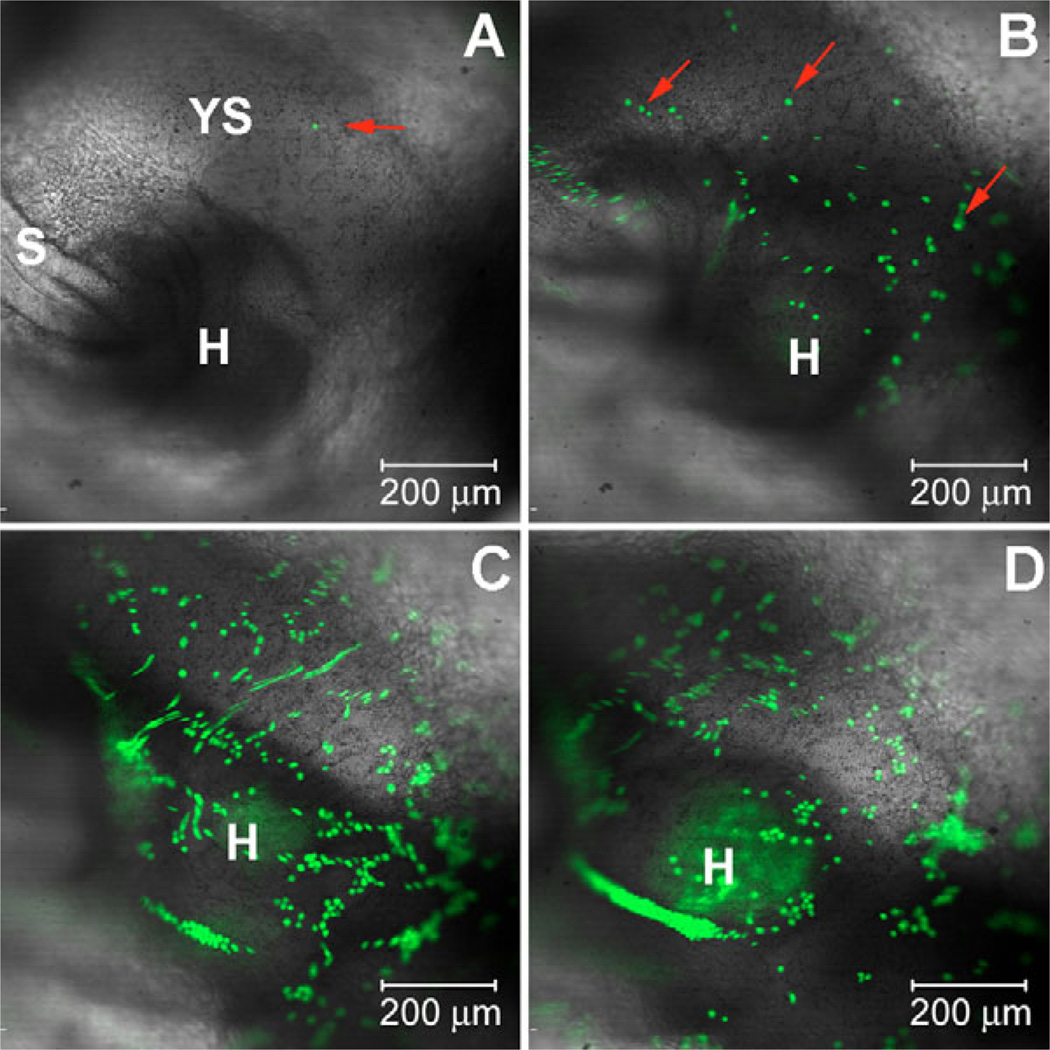
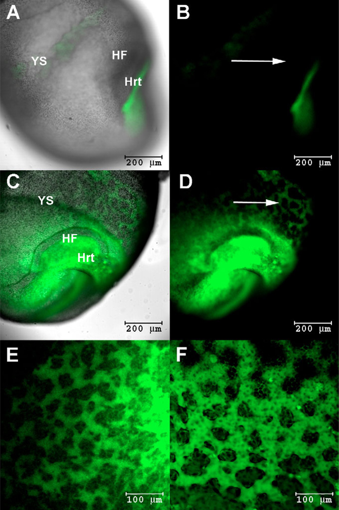
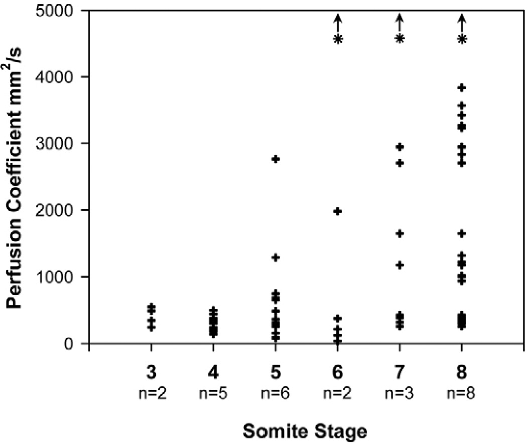
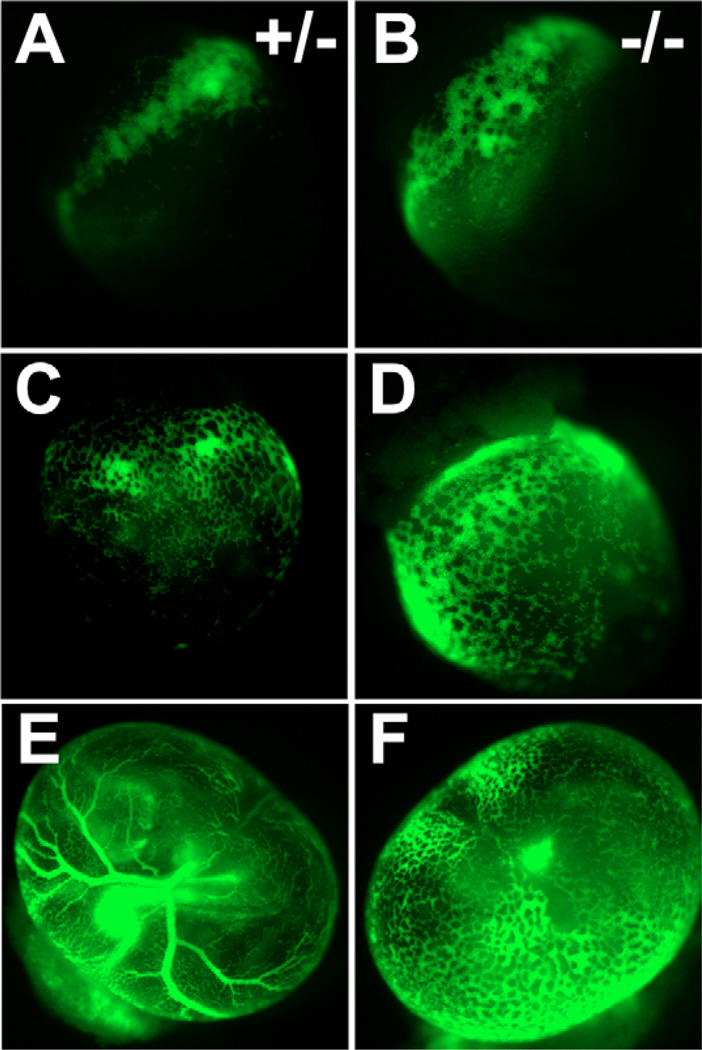
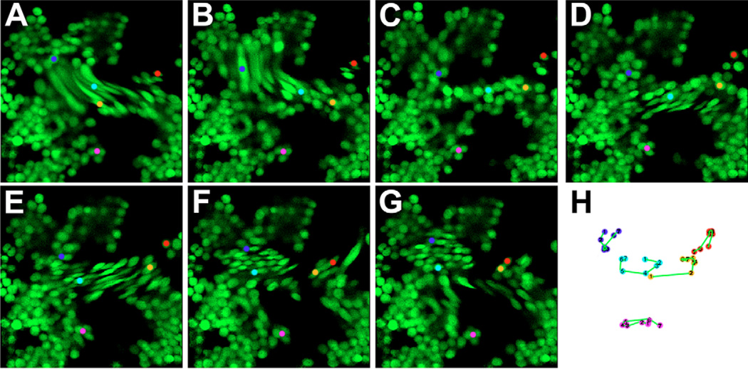
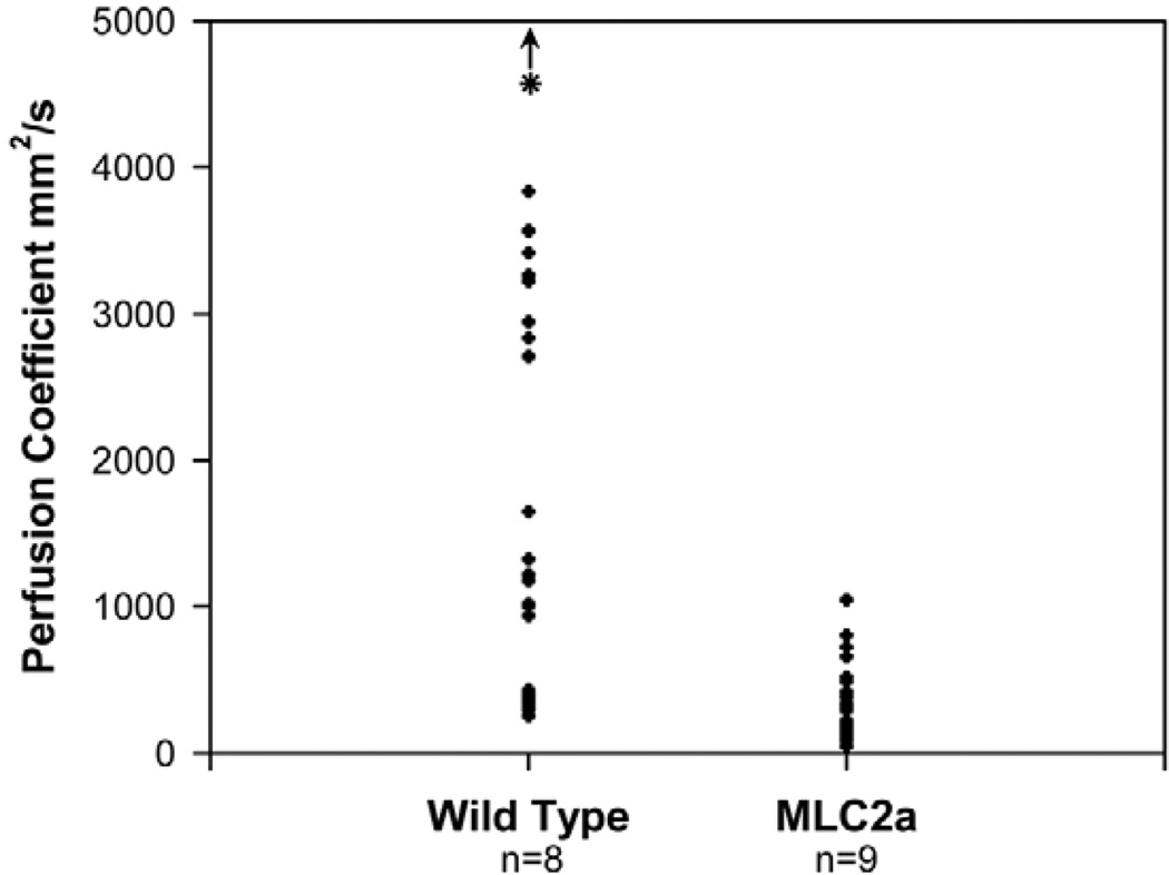
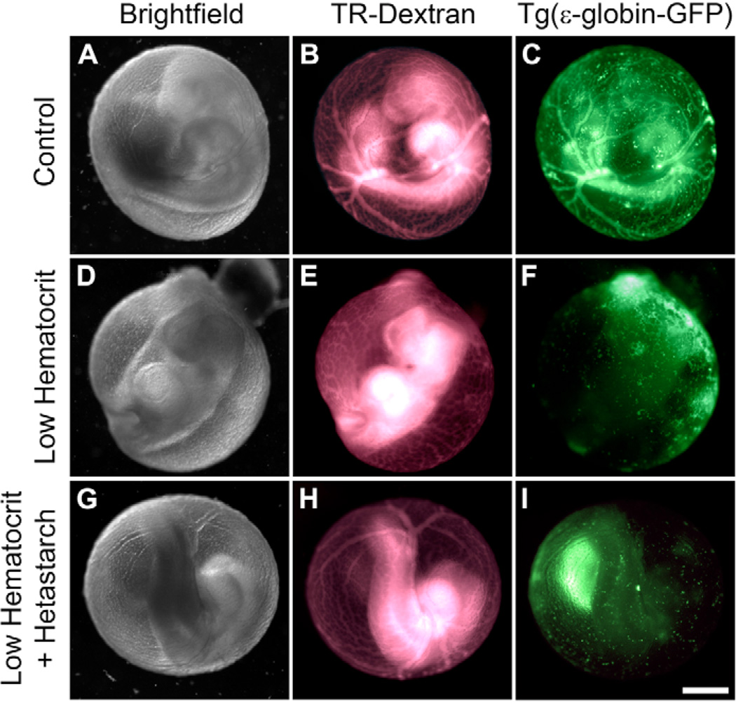
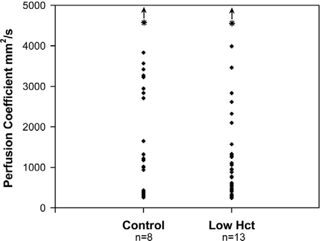
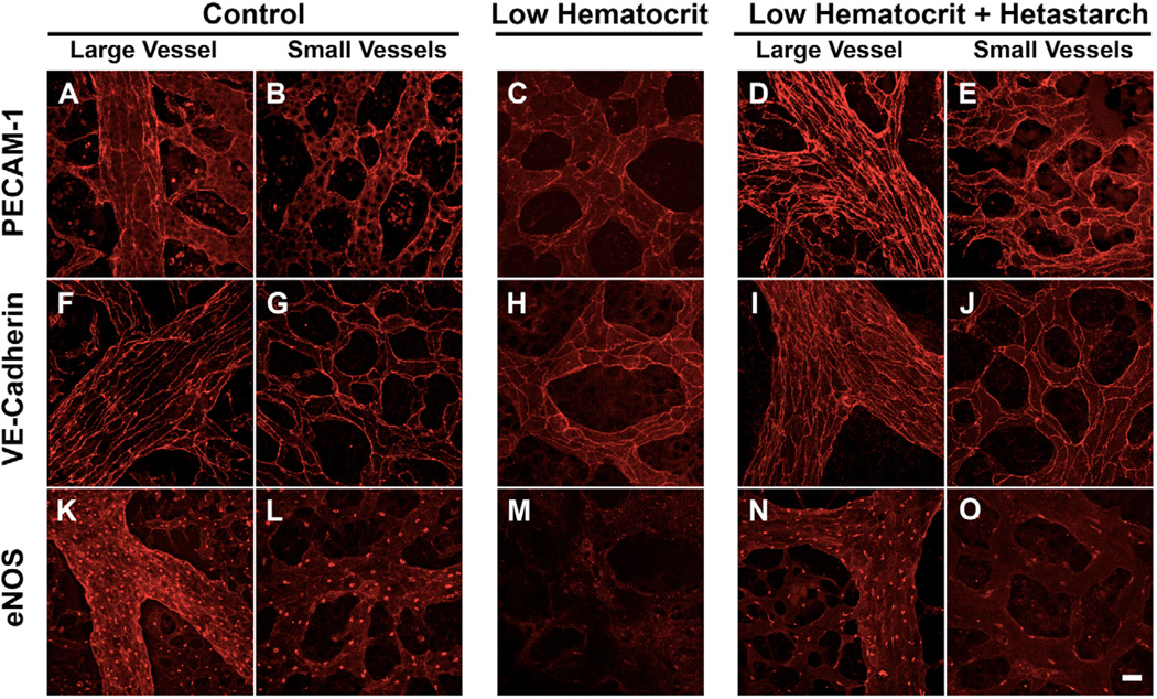
The embryonic heart and vessels are dynamic and form and remodel while functional. Much has been learned about the genetic mechanisms underlying the development of the cardiovascular system, but we are just beginning to understand how changes in heart and vessel structure are influenced by hemodynamic forces such as shear stress. Recent work has shown that vessel remodeling in the mouse yolk sac is secondarily effected when cardiac function is reduced or absent. These findings indicate that proper circulation is required for vessel remodeling, but have not defined whether the role of circulation is to provide mechanical cues, to deliver oxygen or to circulate signaling molecules. Here, we used time-lapse confocal microscopy to determine the role of fluid-derived forces in vessel remodeling in the developing murine yolk sac. Novel methods were used to characterize flows in normal embryos and in embryos with impaired contractility (Mlc2a(-/-)). We found abnormal plasma and erythroblast circulation in these embryos, which led us to hypothesize that the entry of erythroblasts into circulation is a key event in triggering vessel remodeling. We tested this by sequestering erythroblasts in the blood islands, thereby lowering the hematocrit and reducing shear stress, and found that vessel remodeling and the expression of eNOS (Nos3) depends on erythroblast flow. Further, we rescued remodeling defects and eNOS expression in low-hematocrit embryos by restoring the viscosity of the blood. These data show that hemodynamic force is necessary and sufficient to induce vessel remodeling in the mammalian yolk sac.


Figures

Fig. 1
Fig. 1. Time-lapse analysis of the initiation of erythroblast circulation
Images were taken of a 6-somite mouse Tg(ε-globin-GFP) embryo every 6 minutes for a total of 12.1 hours (see Movie 1 in the supplementary material). The yolk sac (YS), heart (H) and somites (S) are indicated. (A) Very few erythroblasts (arrow) are visible in the yolk sac and embryo at the start of the movie (t=0). (B) The same field of view 1 hour later, indicating an increase in erythroblasts. Erythroblasts marked with red arrows are not circulating. (C) Image taken 8.1 hours after the start of the movie, showing more cells moving into the embryo and beginning to fill the heart (H). (D) Fluorescent cells become more evident in the heart towards the end of the time period by 11.7 hours in culture. Clumps of erythroblasts are seen as some cells stop circulating, whereas others continue to move freely.
Fig. 2
Fig. 2. Initiation of plasma flow
10×103 Mr fluorescent dextran was injected into the heart (Hrt) of early mouse embryos. The embryos were incubated for 10 minutes and images were taken at 5× magnification. The presence of fluorescence in the yolk sac after such a short incubation was interpreted as the result of flowing blood plasma. In most 2-somite embryos (5 out of 6 injected embryos), dextran remains localized to the heart (A and B; arrow in B). In one case, however, fluorescent dextran could be observed throughout the yolk sac (ys) (C and D; arrow in D). Plasma circulation is consistently observed after the 3-somite stage (E, 3 somites; F, 6 somites). A and C are images taken with brightfield illumination overlaid with fluorescence images of fluorescein-dextran within the vessels (fluorescent microangiographs); B,D,E and F are microangiographs.
Fig. 3
Fig. 3. Perfusion coefficients of wild-type mouse embryos
Plasma flow magnitude in the early capillary network was determined between the 3- and 8-somite stages by calculating a perfusion coefficient using FRAP. Each measurement is plotted and three measurements were made from each embryo. The total number of embryos analyzed is indicated for each stage. The upper range of perfusion coefficients increases as cardiogenesis progresses and at the 6-somite stage and later, flows that are too fast to measure are observed (asterisk and arrow). We observed a range of values even within a given embryo because flow naturally varies throughout the highly branched plexus.
Fig. 4
Fig. 4. Phenotype of Mlc2a−/− mouse embryos
Heterozygous (A,C,E) and knockout (B,D,F) littermates at the 7-somite (A,B), 10- somite (C,D) and 23-somite (9.5 dpc) (E,F) stage. The capillary plexus is demarcated by GFP-expressing erythroblasts. At the 7-somite stage, blood islands have clearly formed in both wild-type (A) and Mlc2a-null (B) embryos and erythroblasts begin to circulate in both wild-type (C) and Mlc2a mutant (D) embryos. However, the plexus retains an immature phenotype and fails to remodel by embryonic day 9.5 in the mutant embryos (F), as compared with wild type (E).
Fig. 5
Fig. 5. Erythroblast motion in Mlc2a−/− mouse embryos
The motion of erythroblasts (green) within vessels of Mlc2a−/− embryos was imaged at two frames/second. Images were taken at (A) 0, (B) 2.5, (C) 5, (D) 7.5, (E) 10, (F) 12.5 and (G) 15 seconds. The motion of individual erythroblasts, marked by colored dots in A–G, were tracked and these tracks are shown (H), illustrating that erythroblasts oscillate with as much retrograde motion as anterograde.
Fig. 6
Fig. 6. Perfusion coefficients in wild-type and Mlc2a−/− mouse embryos
FRAP was used to measure perfusion coefficients within the early embryonic blood vessels in Mlc2a−/− and wild-type embryos at the 8- to 9-somite stage. Each measurement is plotted and three measurements were made from each embryo. The total number of embryos analyzed is indicated for each genotype. For reproducibility, measurements were always taken on the arterial side of the yolk sac near the caudal end of the embryo. Perfusion coefficient ranges were significantly lower in mutant than in wild-type embryos. In wild-type embryos, some perfusion coefficients were too fast to measure using FRAP (asterisk and arrow). By contrast, perfusion coefficients could always be measured in mutant embryos. Perfusion coefficients up to 3835 µm2/second were found in wild-type embryos, whereas the maximum perfusion rate seen in Mlc2a mutant embryos was 1045 µm2/second.
Fig. 7
Fig. 7. Plasma viscosity alters yolk sac remodeling
Control, low-hematocrit and low-hematocrit+hetastarch mouse embryos were prepared as described (see Materials and methods). Brightfield images show that control embryos turned (A) and yolk sac vessels remodeled into a hierarchical, branched phenotype as evident from Texas Red (TR)-dextran microangiograms (B). eGFP expression (green) from the Tg(ε-globin-GFP) construct shows that erythroblasts are evident in all parts of the yolk sac vasculature (C). The embryo shown in A–C was given a score of 5 for both turning and remodeling. Embryos with sequestered erythroblasts often did not turn but continued to develop after 24 hours in culture (D). The yolk sac vasculature did not remodel and retained features of an immature plexus (E). Erythroblasts remained confined to the blood islands (F). This embryo was scored a 1 for both turning and remodeling. Embryo turning was restored (score=5) (G) and yolk sac remodeling was rescued (score=5) (H) in embryos with sequestered erythroblasts (I) after injection of the hetastarch solution. Scale bar: 500 µm.
Fig. 8
Fig. 8. Perfusion coefficients in low-hematocrit mouse embryos
To ensure that plexus plasma flow was not hampered by immobilizing the erythroblasts to the yolk sac, we determined the perfusion coefficient range in wild-type and acrylamide-treated embryos at 8- to 9-somites. Each measurement is plotted and three measurements were made from each embryo. The total number of embryos analyzed is indicated for each treatment. A similar range of measurements is seen in control and acrylamide-treated embryos indicating that plasma flow is comparable between these two groups.
Fig. 9
Fig. 9. Endothelial cell immunohistochemisty
Confocal images of mouse yolk sac vessels stained with PECAM-1 (A–E), VE-cadherin (F–J), and eNOS (K–O). A,F,K show large vessels from control yolk sacs; B,G,L are images of small vessels in controls. C,H,M are images of immunostaining in small, unremodeled vessels in low-hematocrit embryos, whereas D,I,N and E,J,O are images of large and small vessels, respectively, in low-hematocrit+ hetastarch embryos. A noticeable reduction in eNOS staining was seen in low-hematocrit embryos, but both VE-cadherin and PECAM-1 staining persisted and remained localized to the plasma membrane. Elongated endothelial cell morphology in larger vessels is seen when viscous circulation is present, but unremodeled vessels had a more rounded morphology. Scale bar: 20 µm.

Similar articles

Cited by

References

    1. Akers W, Haidekker MA. A molecular rotor as viscosity sensor in aqueous colloid solutions. J. Biomech. Eng. 2004;126:340–345. - PubMed
    1. Argraves WS, Drake CJ. Genes critical to vasculogenesis as defined by systematic analysis of vascular defects in knockout mice. Anat. Rec. A Discov. Mol. Cell. Evol. Biol. 2005;286:875–884. - PubMed
    1. Armulik A, Abramsson A, Betsholtz C. Endothelial/pericyte interactions. Circ. Res. 2005;97:512–523. - PubMed
    1. Carmeliet P, Lampugnani MG, Moons L, Breviario F, Compernolle V, Bono F, Balconi G, Spagnuolo R, Oostuyse B, Dewerchin M, et al. Targeted deficiency or cytosolic truncation of the VE-cadherin gene in mice impairs VEGF-mediated endothelial survival and angiogenesis. Cell. 1999;98:147–157. - PubMed
    1. Cheng C, van Haperen R, de Waard M, van Damme LC, Tempel D, Hanemaaijer L, van Cappellen GW, Bos J, Slager CJ, Duncker DJ, et al. Shear stress affects the intracellular distribution of eNOS: direct demonstration by a novel in vivo technique. Blood. 2005;106:3691–3698. - PubMed

Publication types