. 2007;35(17):5861-73.
doi: 10.1093/nar/gkm637. Epub 2007 Aug 24.

CTnDOT integrase performs ordered homology-dependent and homology-independent strand exchanges

Affiliations

CTnDOT integrase performs ordered homology-dependent and homology-independent strand exchanges

Karolina Malanowska et al. Nucleic Acids Res. 2007.

Abstract

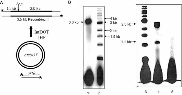
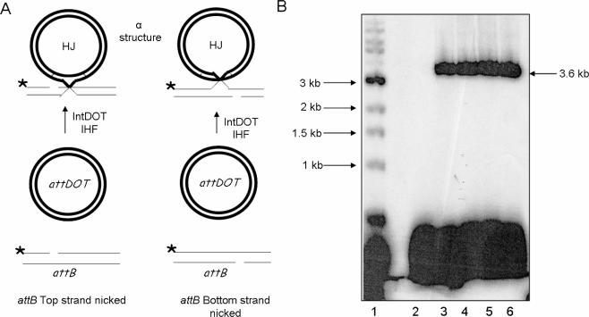
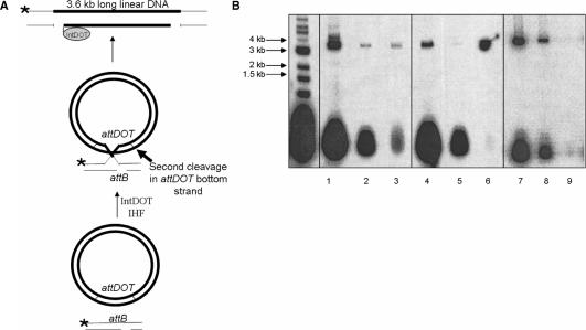
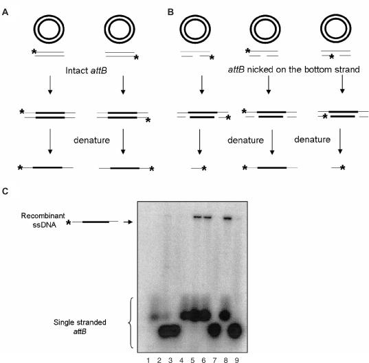
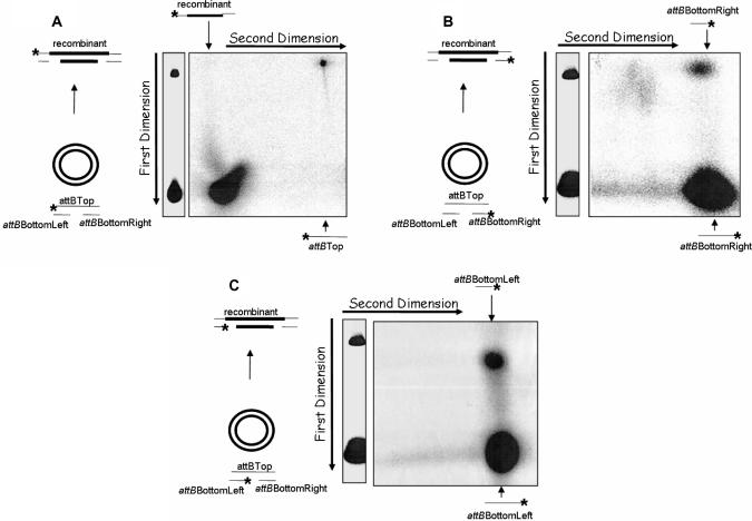
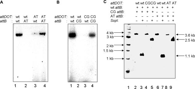
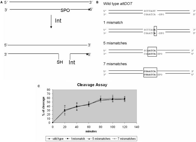
Although the integrase (IntDOT) of the Bacteroides conjugative transposon CTnDOT has been classified as a member of the tyrosine recombinase family, the reaction it catalyzes appears to differ in some features from reactions catalyzed by other tyrosine recombinases. We tested the ability of IntDOT to cleave and ligate activated attDOT substrates in the presence of mismatches. Unlike other tyrosine recombinases, the results revealed that IntDOT is able to perform ligation reactions even when all the bases within the crossover region are mispaired. We also show that there is a strong bias in the order of strand exchanges during integrative recombination. The top strands are exchanged first in reactions that appear to require 2 bp of homology between the partner sites adjacent to the sites of cleavage. The bottom strands are exchanged next in reactions that do not require homology between the partner sites. This mode of coordination of strand exchanges is unique among tyrosine recombinases.


Figures

Figure 1.
Figure 1.
Restriction digest analysis of IntDOT recombinant. (A) The recombination reaction mediated by IntDOT occurs in the presence of E. coli IHF. The reaction occurs between a linear radiolabeled (asterisk) attB DNA and an attDOT site cloned on a plasmid. The product of the reaction is a linear 3.6 kb long recombinant. Cleavage by SspI should generate fragments of 1.1 and 2.5 kb. (B) Agarose gel analysis of recombination products. Lane 1, 3.6 kb long linear recombinant. Lane 2, 1 kb ladder. Lane 3, 1 kb ladder. Lane 4, products of the recombination reaction digested with SspI enzyme. Lane 5, the recombination reaction with no IntDOT and IHF added.
Figure 2.
Figure 2.
Recombination reactions with attB suicide substrates nicked on either the top or bottom DNA strand. (A) Recombination reactions with attB nicked suicide substrates. The attB substrates contain a nick at the site of cleavage in the top (left) or bottom (right) strands. Strand exchange of an attB substrate with a nick in the top strand occurs if the bottom strands are exchanged first (left). If the nick is in the bottom strand of attB, a strand exchange occurs if the top strand is exchanged first (right). The strand with a nick placed in a cleavage site should not undergo recombination. The HJ intermediate has a form of an α structure. (B) Agarose gel analysis of recombination reactions with attB substrates that contain a ssDNA nick at the IntDOT cleavage site in the top or in the bottom DNA strands. Lane: 1, 1 kb ladder. Lanes 2 and 3 are reactions with a nick in the top (lane 2) or bottom (lane 3) attB strand. Lane 4 is a reaction with an intact attB. Lanes 5 and 6 are attB substrates that contained a nick in the top (lane 5) or bottom (lane 6) strand that were treated with T4 DNA ligase to seal the nick.
Figure 3.
Figure 3.
Restriction digest analysis of the product of the IntDOT-mediated recombination reaction with suicide attB. Recombination reaction with an intact attB substrate labeled at both ends (lane 2) and digested with SspI (lane 1). Reactions with bottom strand nicked attB (lane 4) and digested with SspI (lane 3). Molecular weight standards are in lane 5.
Figure 4.
Figure 4.
Isolation of covalent IntDOT/DNA complexes, using the SDS/KCl precipitation method. (A) Recombination reaction with an attB substrate nicked in the bottom DNA strand. After formation of the HJ, a second DNA cleavage occurs in the attDOT site in the bottom DNA strand that is not followed by DNA ligation. The product of this reaction is a linear 3.6 kb long DNA fragment containing two nicks in the bottom DNA strand and protein attached to the 3′ attDOT DNA end. (B) Recombination reaction with intact attB substrates (lanes 1–3), with attB containing a nick in IntDOT cleavage site in the bottom DNA strands (lanes 4–6) and with attB containing a nick in IntDOT cleavage site on the bottom DNA strand where proteinase K was added before SDS/KCl precipitation (lanes 7–9). Lanes 1, 4 and 7 contain untreated reactions. Lanes 2, 5 and 8 contain supernatants after SDS/KCl precipitation. Lanes 3, 6 and 9 contain pellets after SDS/KCl precipitation.
Figure 5.
Figure 5.
Denaturing agarose gel analysis of products.(A) Predicted products of recombination reactions with intact attB. (B) Predicted products of recombination reactions with suicide attB. (C) Denaturing agarose gel analysis of intact attB reaction with either attB strand labeled yields intact a 3.6 kb single-stranded DNA. The product of a strand exchange with a suicide attB substrate with the top strand labeled should yield a 3.6 kb single-stranded DNA. If either of the bottom strands is labeled, a 40-base single-stranded DNA should be generated. Denatured substrates: lane 1, denatured top strand of intact attB; lane 2, denatured left, bottom strand of attB; lane 3, denatured right, bottom strand of attB; lane 4, denatured bottom strand of intact attB. Recombination reactions: lane 5, with intact attB with the top strand labeled; lane 6, with intact attB with the bottom strand labeled, lane 7, with suicide attB with the bottom right strand labeled; lane 8, with suicide attB with the top strand labeled; lane 9, with suicide attB with the bottom left strand labeled.
Figure 6.
Figure 6.
Two-dimensional gel analysis of the products generated by IntDOT-mediated recombination. The attB substrates contain a nick in the IntDOT cleavage site in the bottom DNA strand. attB DNA was radiolabeled at one 5′ end (asterisk), incubated with attDOT DNA in a standard recombination reaction and treated as described in Materials and Methods section. (A) The top attB DNA strand labeled. (B) The right bottom strand (attBBottomRight oligo) labeled. (C) The left bottom attB strand (attBBottomLeft oligo) labeled.
Figure 7.
Figure 7.
IntDOT recombination reactions with mutated attB and attDOT substrates. Mutations were introduced in the 2 bp region of homology in the left side of IntDOT crossover region. The GC sequence was changed to AT or CG, respectively. (A) Recombination between sites with GC to AT mutations. Lane 1, wild-type attDOT and wild-type attB; lane 2, wild-type attDOT and attBAT; lane 3, wild-type attB and attDOTAT; lane 4, attDOTAT and attBAT. (B) Recombination between sites with GC to CG mutations. Lane 1, wild-type attDOT and wild-type attB; lane 2, wild-type attDOT and attBCG; lane 3, wild-type attB and attDOTCG; lane 4, attDOTCG and attBCG. (C) A competition assay. Equimolar concentrations of wild-type attB labeled on the top strand and mutated attB labeled on the bottom strand were added to the reaction with wild-type attDOT or mutated attDOT, respectively. Reactions were digested with SspI enzyme. Digestion of the recombinant with a top strand labeled should give 1.1 kb DNA fragment. Digestion of the recombinant with the bottom strand labeled should give 2.5 kb fragment.
Figure 8.
Figure 8.
Phosphorothioate cleavage assay. (A) The labeled attDOT DNA substrate (asterisk) contains a 5′-bridging phosphorothioate linkage at the site of cleavage. If the enzyme cleaves the DNA, it becomes covalently bound to the substrate DNA. This reaction is irreversible. (B) The attDOT substrates used in cleavage assays. (C) Results of the phosphorothioate cleavage assay with wild-type attDOT sequence and attDOT sequence containing 1, 5 and 7 missmatches within the crossover region. The reaction efficiencies were calculated as described in Materials and Methods section.
Figure 9.
Figure 9.
Ligation assay. (A) The labeled DNA substrate (asterisk) contains a p-nitrophenol linkage incorporated at the 3′ site of cleavage, which mimics phosphotyrosyl bond between IntDOT and DNA. This substrate can be recognized and ligated by λ or CTnDOT integrase. (B) attDOT and attP substrates used in ligation assays. (C) Results of ligation assays with variations of attDOT (left) and λ attP (right). The reaction efficiencies were calculated as described in Materials and Methods section.

Similar articles

Cited by

References

    1. Salyers AA, Shipman JA. 11: Getting in touch with your prokaryotic self: mammal-microbe interactions. In: Staley JT, Reysenbach A.-L, editors. Biodiversity of Microbial Life. Wiley-Liss, Inc.; 2002. pp. 315–341.
    1. Macy JM, Probst I. The biology of gastrointestinal bacteroides. Annu. Rev. Microbiol. 1979;33:561–594. - PubMed
    1. Shoemaker NB, Vlamakis H, Hayes K, Salyers AA. Evidence for extensive resistance gene transfer among Bacteroides spp and between Bacteroides and other genera in the human colon. Appl. Environ. Microbiol. 2001;67:561–568. - PMC - PubMed
    1. Whittle G, Shoemaker NB, Salyers AA. The role of Bacteroides conjugative transposons in the dissemination of antibiotic resistance genes. Cell. Mol. Life Sci. 2002;59:2044–2054. - PMC - PubMed
    1. Cheng Q, Paszkiet BJ, Shoemaker NB, Gardner JF, Salyers AA. Integration and excision of a Bacteroides conjugative transposon, CTnDOT. J. Bacteriol. 2000;182:4035–4043. - PMC - PubMed

Publication types