Identification of eukaryotic promoter regulatory elements using nonhomologous random recombination
- PMID: 17720707
- PMCID: PMC2034452
- DOI: 10.1093/nar/gkm634
Identification of eukaryotic promoter regulatory elements using nonhomologous random recombination
Abstract
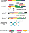
Understanding the regulatory logic of a eukaryotic promoter requires the elucidation of the regulatory elements within that promoter. Current experimental or computational methods to discover regulatory motifs within a promoter can be labor intensive and may miss redundant, unprecedented or weakly activating elements. We have developed an unbiased combinatorial approach to rapidly identify new upstream activating sequences (UASs) in a promoter. This approach couples nonhomologous random recombination with an in vivo screen to efficiently identify UASs and does not rely on preconceived hypotheses about promoter regulation or on similarity to known activating sequences. We validated this method using the unfolded protein response (UPR) in yeast and were able to identify both known and potentially novel UASs involved in the UPR. One of the new UASs discovered using this approach implicates Crz1 as a possible activator of Hac1, a transcription factor involved in the UPR. This method has several advantages over existing methods for UAS discovery including its speed, potential generality, sensitivity and lack of false positives and negatives.
Figures


Similar articles
-
Gcn4p and novel upstream activating sequences regulate targets of the unfolded protein response.PLoS Biol. 2004 Aug;2(8):E246. doi: 10.1371/journal.pbio.0020246. Epub 2004 Aug 17. PLoS Biol. 2004. PMID: 15314660 Free PMC article.
-
Autoregulation of the HAC1 gene is required for sustained activation of the yeast unfolded protein response.Genes Cells. 2004 Feb;9(2):95-104. doi: 10.1111/j.1365-2443.2004.00704.x. Genes Cells. 2004. PMID: 15009095
-
Expression of the INO2 regulatory gene of Saccharomyces cerevisiae is controlled by positive and negative promoter elements and an upstream open reading frame.Mol Microbiol. 2001 Mar;39(5):1395-405. Mol Microbiol. 2001. PMID: 11251853
-
ATF/CREB sites present in sub-telomeric regions of Saccharomyces cerevisiae chromosomes are part of promoters and act as UAS/URS of highly conserved COS genes.J Mol Biol. 2002 May 31;319(2):407-20. doi: 10.1016/S0022-2836(02)00322-4. J Mol Biol. 2002. PMID: 12051917
-
Bax induces activation of the unfolded protein response by inducing HAC1 mRNA splicing in Saccharomyces cerevisiae.Yeast. 2012 Sep;29(9):395-406. doi: 10.1002/yea.2918. Epub 2012 Aug 18. Yeast. 2012. PMID: 22903551
Cited by
-
Tailored UPRE2 variants for dynamic gene regulation in yeast.Proc Natl Acad Sci U S A. 2024 May 7;121(19):e2315729121. doi: 10.1073/pnas.2315729121. Epub 2024 Apr 30. Proc Natl Acad Sci U S A. 2024. PMID: 38687789 Free PMC article.
-
Feature Identification of Compensatory Gene Pairs without Sequence Homology in Yeast.Comp Funct Genomics. 2012;2012:653174. doi: 10.1155/2012/653174. Epub 2012 Aug 16. Comp Funct Genomics. 2012. PMID: 22952430 Free PMC article.
-
Tracking Effects of SIL1 Increase: Taking a Closer Look Beyond the Consequences of Elevated Expression Level.Mol Neurobiol. 2018 Mar;55(3):2524-2546. doi: 10.1007/s12035-017-0494-6. Epub 2017 Apr 11. Mol Neurobiol. 2018. PMID: 28401474
-
Transcriptional regulation in Saccharomyces cerevisiae: transcription factor regulation and function, mechanisms of initiation, and roles of activators and coactivators.Genetics. 2011 Nov;189(3):705-36. doi: 10.1534/genetics.111.127019. Genetics. 2011. PMID: 22084422 Free PMC article. Review.
-
Genome-wide and molecular evolution analysis of the Poplar KT/HAK/KUP potassium transporter gene family.Ecol Evol. 2012 Aug;2(8):1996-2004. doi: 10.1002/ece3.299. Epub 2012 Jul 19. Ecol Evol. 2012. PMID: 22957200 Free PMC article.
References
-
- Srikanth CV, Vats P, Bourbouloux A, Delrot S, Bachhawat AK. Multiple cis-regulatory elements and the yeast sulphur regulatory network are required for the regulation of the yeast glutathione transporter, Hgt1p. Curr. Genet. 2005;47:345–358. - PubMed
Publication types
MeSH terms
Substances
Grants and funding
LinkOut - more resources
Full Text Sources
Other Literature Sources
Molecular Biology Databases

