. 2007;35(17):5809-18.
doi: 10.1093/nar/gkm613. Epub 2007 Aug 24.

The yeast Pif1p DNA helicase preferentially unwinds RNA DNA substrates

Affiliations

The yeast Pif1p DNA helicase preferentially unwinds RNA DNA substrates

Jean-Baptiste Boulé et al. Nucleic Acids Res. 2007.

Abstract

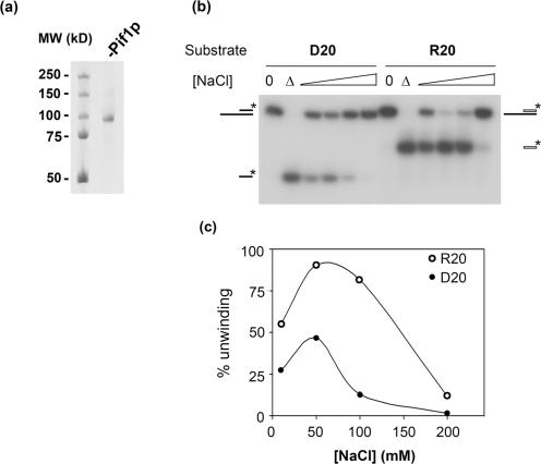
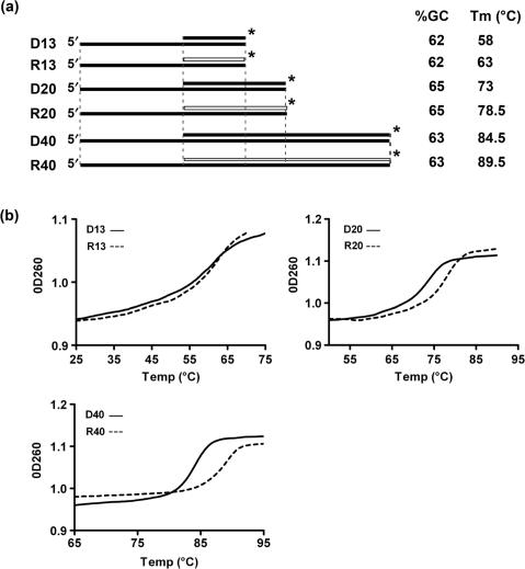
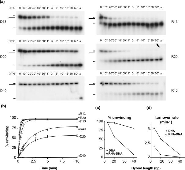
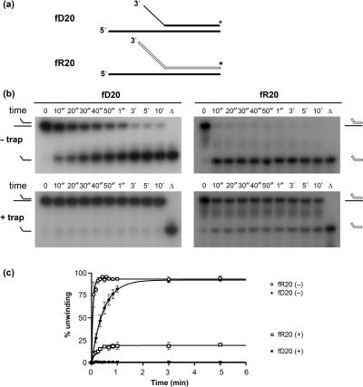
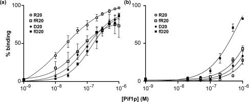
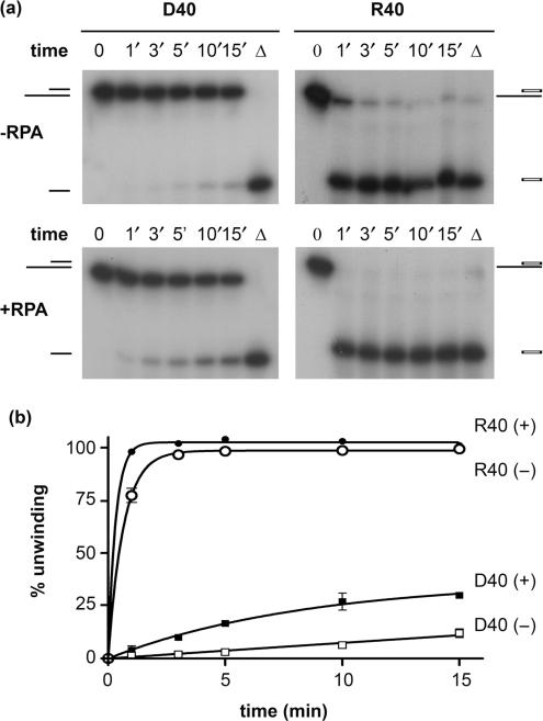
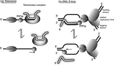
Pif1p is the prototypical member of the PIF1 family of DNA helicases, a subfamily of SFI helicases conserved from yeast to humans. Baker's yeast Pif1p is involved in the maintenance of mitochondrial, ribosomal and telomeric DNA and may also have a general role in chromosomal replication by affecting Okazaki fragment maturation. Here we investigate the substrate preferences for Pif1p. The enzyme was preferentially active on RNA-DNA hybrids, as seen by faster unwinding rates on RNA-DNA hybrids compared to DNA-DNA hybrids. When using forked substrates, which have been shown previously to stimulate the enzyme, Pif1p demonstrated a preference for RNA-DNA hybrids. This preferential unwinding could not be correlated to preferential binding of Pif1p to the substrates that were the most readily unwound. Although the addition of the single-strand DNA-binding protein replication protein A (RPA) stimulated the helicase reaction on all substrates, it did not diminish the preference of Pif1p for RNA-DNA substrates. Thus, forked RNA-DNA substrates are the favored substrates for Pif1p in vitro. We discuss these findings in terms of the known biological roles of the enzyme.


Figures

Figure 1.
Figure 1.
Pif1p unwinding activity as a function of NaCl concentration. (a) Coomassie gel staining of the purified recombinant nuclear isoform of Pif1p. (b) Unwinding of DNA–DNA (D20, left) and RNA–DNA (R20, right) hybrids at increasing salt concentrations. A total of 60 nM Pif1p was incubated with 1 nM D20 or R20 substrates at 35°C for 15 min. Products of the reaction were analyzed as described in the Materials and Methods section. (0) indicates zero time point, (▵) indicates heat denatured sample. (c) Quantification of the unwinding of RNA–DNA (R20, open circles) and DNA–DNA (D20, closed circles) substrates shown in (b). This experiment was done several times with similar results.
Figure 2.
Figure 2.
Structure and stability of helicase substrates. (a) Schematic of the structures of substrates used for helicase assays. Black bars represent DNA strands; white bars are RNA strands. Vertical dotted lines indicate regions of identical sequence among the various substrates. The asterisk indicates the position of the 32P label. GC content of the substrates in the hybrid region and the experimentally determined Tm (±0.5°C, as in b) are also indicated for each substrate. (b) Thermal denaturation profiles of the helicase substrates depicted in (a). The experiment was done at least twice for each substrate with identical results. A representative experiment is shown.
Figure 3.
Figure 3.
Kinetics of unwinding by Pif1p of RNA–DNA and DNA–DNA hybrids of various sizes. (a) Unwinding by Pif1p of the substrates D13, D20, D40 and their RNA–DNA hybrid counterparts during a 1-h time course. For these experiments, 60 nM Pif1p was incubated with 1 nM substrate. Aliquots were removed at the indicated time points and analyzed on non-denaturing PAGE gels as described in the Materials and Methods section. (b) Quantification of RNA–DNA and DNA–DNA substrate unwinding by Pif1p. Errors bars indicate SD. Amplitude (% unwinding) of the reaction and apparent turnover rate for the different substrates are as follows A(D13) = 96 ± 2%; k(D13) = 2 ± 0.15 min−1; A(R13) = 97 ± 0.4%; k(R13) = 5 ± 0.15 min−1; A(D20) = 57 ± 1%; k(D20) = 0.5 ± 0.03 min−1; A(R20) = 99 ± 1%; k(R20) = 3 ± 0.2 min−1; A(D40) = 8 ± 1%; k(D40) = 0.02 ± 0.005 min−1; A(R40) = 80 ± 1%; k(R40) = 0.5 ± 0.02 min−1. (c) % unwinding and (d) turnover rate of the helicase reactions as a function of the hybrid length, derived from the three independent experiments analyzed in (b). SD are displayed but are too small to be visible at this scale.
Figure 4.
Figure 4.
Activity of Pif1p on forked substrates at 25°C. (a) Structure of the forked substrates, using the same conventions as in Figure 2a. (b) Unwinding of fD20 and fR20 forked substrates in multiple cycle (– trap) and single cycle conditions (+ trap). (c) Quantification of three independent unwinding reactions of the fD20 and fR20 forked substrates. (+) and (–) indicate, respectively, reactions done in the presence and absence of a single-strand DNA trap. Amplitude and apparent turnover rates are as follows. Multiple cycle conditions: A(fD20) = 92 ± 2%, k(fD20) = 2.1 ± 0.3 min−1; A(fR20) = 93 ± 1%, k(fR20) = 18.3 ± 2 min−1. For single cycle kinetics, no unwinding was observed for the fD20 substrate; values for fR20 were A(fR20) = 19 ± 1%, k(fR20) = 5.2 ± 0.7 min−1.
Figure 5.
Figure 5.
Pif1p binding to forked and unforked substrates. Increasing amounts of Pif1p were incubated at 25°C with 50 pM nucleic acid substrate, in absence (a) or presence (b) of the nonhydrolyzable nucleotide analog AMPPNP, and analyzed on 12% non-denaturing gels as described in the Materials and Methods section. Data from three different experiments were averaged and plotted. Error bars represent the SD between the three experiments. Binding parameters are as follows; in the absence of a nucleotide, Kd(D20) = 1.0 × 10⁻⁷ M; Kd(R20) = 1.7 × 10⁻⁸ M; Kd(fD20) = 1.3 × 10⁻⁷ M; Kd(fR20) = 5.2 × 10⁻⁸ M; in the presence of AMPPNP, Kd(D20) = 2.0 × 10⁻⁶ M; Kd(R20) = 3.2 × 10⁻⁶ M; Kd(fD20) = 3.2 × 10⁻⁷ M; Kd(fR20) = 1.4 × 10⁻⁶ M.
Figure 6.
Figure 6.
Pif1p activity is stimulated by yeast RPA. (a) Unwinding of the D40 and R40 substrates in presence or absence of yeast RPA. 100 nM Pif1p was incubated with 1 nM nucleic acid substrate at 25°C, in the presence or absence of 50 ng/µl recombinant yeast RPA (generous gift from P. Sung). (b) Quantification of three independent experiments as shown in (a). Bars indicate SDs, which are present on every point. Amplitude and turnover rates are as follows. In the presence of RPA, A(D40) = 37 ± 5%, k(D40) = 0.1 ± 0.02 min−1; A(R40) = 100%, k(R40) = 3 ± 0.2 min−1; in the absence of RPA, A(R40) = 99 ± 1%, k(R40) = 1.5 ± 0.07 min−1. Reaction parameters for D40 were not determined.
Figure 7.
Figure 7.
Pif1p exists as a monomer in solution. Pif1p was subjected to centrifugation through a 15–40% glycerol gradient. The protein markers used as molecular weight standards were Carbonic Anhydrase (29 kDa; s20,w = 2.8 S), BSA (66 kDa, s20,w = 4.41 S), ADH (150 kDa, s20,w = 4.8 S), β-Amylase (200 kDa, s20,w = 8.9 S) and Thyroglobulin (669kDa, s20,w = 19.4 S). Two hundred and fifty micro liter fractions from the 5 ml gradient were analyzed for protein content by Coomassie gel staining to determine elution peak of molecular weight markers, and by anti-His western blotting for the detection of Pif1p.
Figure 8.
Figure 8.
Hypothetical RNA–DNA hybrid substrates for Pif1p. Two possible RNA–DNA hybrids are shown. RNA strands are shown in white, DNA strands in black (a) Pif1p acts catalytically to remove telomerase from DNA ends, possibly by unwinding the RNA–DNA hybrid formed by TLC1 and the end of the telomere (7,8,12). (b) Pif1p could be involved in removing stable RNA–DNA hybrids in front of the replication fork. At the rDNA locus (pictured), Pif1p could help remove rDNA transcripts that approach the RFB established by Fob1p (36). Elsewhere, the R-loop could originate from a transcribed gene or an incompletely processed Okazaki fragment RNA primer (37,38). An alternative hypothesis has been proposed for the role of Pif1p in Okazaki fragment processing that does not involve RNA–DNA hybrid unwinding, in which Pif1p extends the DNA flap in front of the lagging strand polymerase (14).

References

    1. Sharma S, Doherty KM, Brosh R.M., Jr Mechanisms of RecQ helicases in pathways of DNA metabolism and maintenance of genomic stability. Biochem. J. 2006;398:319–337. - PMC - PubMed
    1. Delagoutte E, von Hippel PH. Helicase mechanisms and the coupling of helicases within macromolecular machines. Part II: integration of helicases into cellular processes. Q. Rev. Biophys. 2003;36:1–69. - PubMed
    1. Bessler JB, Torres JZ, Zakian VA. The Pif1p subfamily of helicases: region-specific DNA helicases? Trends Cell. Biol. 2001;11:60–65. - PubMed
    1. Boule JB, Zakian VA. Roles of Pif1-like helicases in the maintenance of genomic stability. Nucleic Acids Res. 2006;34:4147–4153. - PMC - PubMed
    1. Foury F, Kolodynski J. pif mutation blocks recombination between mitochondrial rho+ and rho- genomes having tandemly arrayed repeat units in Saccharomyces cerevisiae. Proc. Natl Acad. Sci. USA. 1983;80:5345–5349. - PMC - PubMed

Publication types

MeSH terms