. 2007;35(17):5789-98.
doi: 10.1093/nar/gkm503. Epub 2007 Aug 24.

Analysis of the XPA and ssDNA-binding surfaces on the central domain of human ERCC1 reveals evidence for subfunctionalization

Affiliations

Analysis of the XPA and ssDNA-binding surfaces on the central domain of human ERCC1 reveals evidence for subfunctionalization

Konstantinos Tripsianes et al. Nucleic Acids Res. 2007.

Abstract

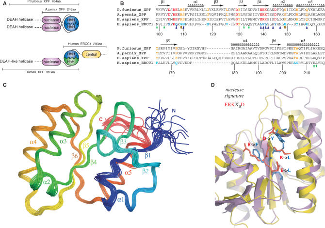
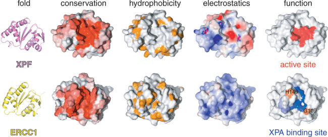
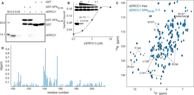
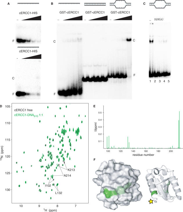
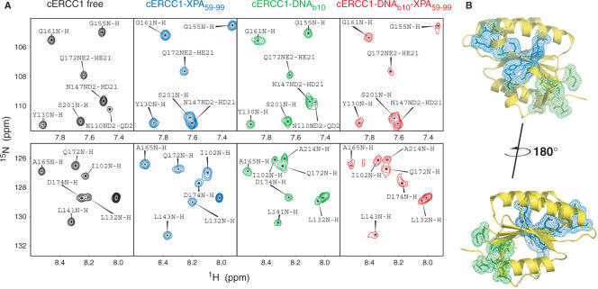
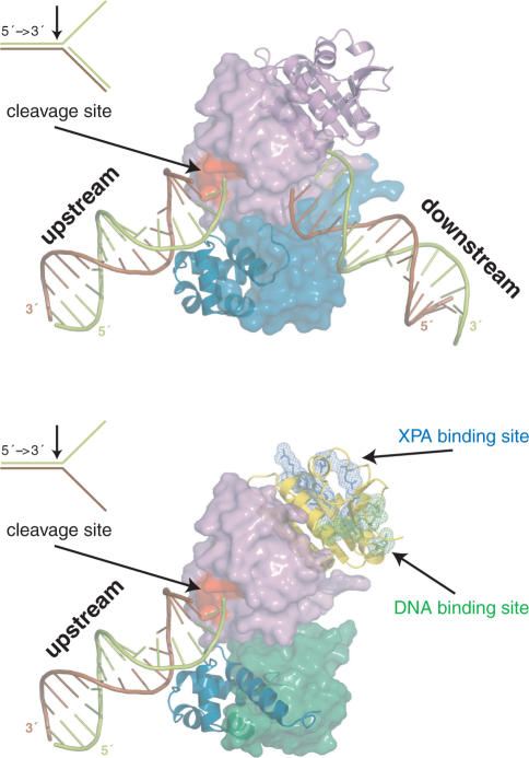
Human ERCC1/XPF is a structure-specific endonuclease involved in multiple DNA repair pathways. We present the solution structure of the non-catalytic ERCC1 central domain. Although this domain shows structural homology with the catalytically active XPF nuclease domain, functional investigation reveals a completely distinct function for the ERCC1 central domain by performing interactions with both XPA and single-stranded DNA. These interactions are non-competitive and can occur simultaneously through distinct interaction surfaces. Interestingly, the XPA binding by ERCC1 and the catalytic function of XPF are dependent on a structurally homologous region of the two proteins. Although these regions are strictly conserved in each protein family, amino acid composition and surface characteristics are distinct. We discuss the possibility that after XPF gene duplication, the redundant ERCC1 central domain acquired novel functions, thereby increasing the fidelity of eukaryotic DNA repair.


Figures

Figure 1.
Figure 1.
(A) Domain organization of the archaeal XPF homodimeric members and the human ERCC1/XPF heterodimer. (B) Structure-based sequence alignment of the nuclease XPF domains from archaea to human and the corresponding central domain of human ERCC1. Secondary structure elements of the prototype XPF nuclease fold are indicated at the top of the sequences. Catalytic residues in the XPF nucleases are colored red and their corresponding substitutions in ERCC1 blue. Other invariant residues in XPF domains are depicted in orange, while the ERCC1 equivalents are depicted in cyan. Residues of cERCC1 perturbed largely upon XPA titration are indicated by blue triangles and those appear only in the final complex by brown ellipses. Green asterisks indicate cERCC1 residues perturbed by DNA titration. cERCC1 sequence is numbered at the bottom. (C) Ensemble of the final 20 structural conformers of cERCC1 as determined by solution NMR. Secondary structure elements and N- and C-termini are labeled. (D) Superposition of the crystal XPF nuclease structure (2bgw) from A. pernix (purple) and the solution NMR structure (2jpd) of human cERCC1 (yellow). Emphasis is given to the nuclease signature and the corresponding substitutions in cERCC1.
Figure 3.
Figure 3.
Common fold, different properties and distinct functions for XPF and ERCC1. Sequence conservation either for XPF or ERCC1 proteins is colored from white (non-conserved) to red (highly conserved). The opposite face has no significant conservation for either protein. Hydrophobic side chains are colored orange. Electrostatic surface potentials were calculated using APBS (39) and colored blue for positive or red for negative charge potential. Active site residues of XPF were chosen based on previous mutational studies (5) and the XPA-binding site of ERCC1 was identified by our NMR titrations.
Figure 2.
Figure 2.
ERCC1–XPA interactions. (A) GST pull-down assay with 3 µg of GST or GST-XPA fusion proteins in the presence or absence of 2 µM cERCC1. Here, 0.2 and 0.04 refer to respectively 20 and 4% of the input cERCC1 protein present in the GST pull-down assay. (B) Semi quantitative GST pull-down assay showing the fraction of cERCC1 bound to 3 µg of GST-XPA at the indicated [cERCC1] (µM). The upper part of inset shows a representative GST pull-down assay, where the various cERCC1 concentrations used are depicted above (10, 3, 1, 0.3 and 0.1 µM). The lower panel shows respectively 100, 20 and 4% of cERCC1 added to the assay. (C) Chemical shift perturbation of the cERCC1 1H-15N HSQC upon complex formation with XPA. Free cERCC1 spectrum is in black and XPA-bound spectrum in blue, while brown circles in the bound spectrum indicate G109 and H149 resonances. (D) Normalized chemical shift changes between free and XPA-bound forms versus the cERCC1 sequence. The p.p.m. difference for G109 and H149 was calculated by their resonances in the free cERCC1 spectrum at pH 5.5, and are colored brown.
Figure 4.
Figure 4.
DNA binding by cERCC1. (A) EMSA with a 20-mer ssDNA substrate using increasing concentrations of cERCC1-HIS protein (0, 0.04, 0.2, 1, and 5 µM), loaded on a 7.5% acrylamide gel (upper panel) or a 3.5% agarose gel (lower panel). (B) EMSA using ssDNA (20-mer), dsDNA (30 bp) and bubble10, a dsDNA with 10 unpaired bases as substrate, in the presence of 0, 0.125, 0.25, 0.5 and 1 µM HIS-GST-cERCC1. (C) Inhibition of cERCC1–b10 DNA complex formation ([cERCC1] = 0.5 µM) by depletion of HIS-GST-cERCC1 from the EMSA reaction by the addition of MagneHis beads (H) or GST agarose beads (G) prior to or after addition of DNA (italic). F: free DNA, C: cERCC1–DNA complex. (D) Chemical shift perturbation after addition of b10 DNA. The spectrum of the free protein is shown in black and the spectrum after addition of equimolar amount of b10 in green. (E) Normalized chemical shift changes between free and b10-bound forms versus the cERCC1 sequence. (F) Surface and cartoon representations of cERCC1 colored according to normalized chemical shifts. Yellow star indicates the position of the C-terminal his-tag that was interfering with DNA binding.
Figure 5.
Figure 5.
Dual function of cERCC1. (A) Indicative portions of the 1H-15N HSQC spectrum for the free cERCC1, bound to either XPA (1:1) or DNA (1:1) separately, and the ternary complex (1:1:1). (B) Views of the two binding sites on cERCC1 structure.
Figure 6.
Figure 6.
Models for the function of the archaeal homodimeric XPF (top) and human ERCC1/XPF heterodimer (bottom) constructed from the structure of the XPF homodimer bound to dsDNA (2bgw), the free structure of ERCC1/XPF C-terminal interacting domains (1z00) and the current structure of the ERCC1 central domain (2jpd). For the homodimer, one protomer is shown in a cartoon and the other in a surface representation. Accordingly, for the heterodimer ERCC1 is in a cartoon and XPF in a surface representation. The protein domains in both cases are colored as in Figure 1A.

References

    1. Nishino T, Morikawa K. Structure and function of nucleases in DNA repair: shape, grip and blade of the DNA scissors. Oncogene. 2002;21:9022–9032. - PubMed
    1. Ciccia A, Ling C, Coulthard R, Yan Z, Xue Y, Meetei AR, Laghmani el H, Joenje H, McDonald N, et al. Identification of FAAP24, a Fanconi anemia core complex protein that interacts with FANCM. Mol. Cell. 2007;25:331–343. - PubMed
    1. Nishino T, Komori K, Ishino Y, Morikawa K. X-ray and biochemical anatomy of an archaeal XPF/Rad1/Mus81 family nuclease: similarity between its endonuclease domain and restriction enzymes. Structure (Camb.) 2003;11:445–457. - PubMed
    1. Heyer WD, Ehmsen KT, Solinger JA. Holliday junctions in the eukaryotic nucleus: resolution in sight? Trends Biochem. Sci. 2003;28:548–557. - PubMed
    1. Enzlin JH, Scharer OD. The active site of the DNA repair endonuclease XPF-ERCC1 forms a highly conserved nuclease motif. EMBO J. 2002;21:2045–2053. - PMC - PubMed

Publication types

MeSH terms