. 2007 Dec 15;93(12):4278-88.
doi: 10.1529/biophysj.107.113043. Epub 2007 Aug 24.

The dynamic orientation of membrane-bound peptides: bridging simulations and experiments

Affiliations

The dynamic orientation of membrane-bound peptides: bridging simulations and experiments

Santi Esteban-Martín et al. Biophys J. .

Abstract

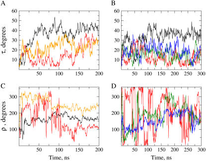
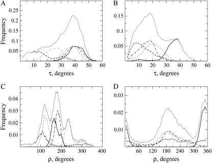
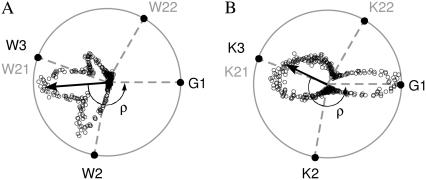
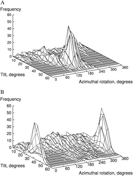
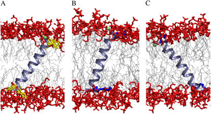
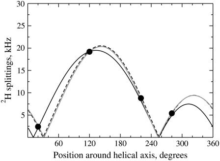
The structural organization in a peptide/membrane supramolecular complex is best described by knowledge of the peptide orientation plus its time-dependent and spatial fluctuations. The static orientation, defined by the peptide tilt and a rotation about its molecular axis, is accessible through a number of spectroscopic methods. However, peptide dynamics, although relevant to understand the functionality of these systems, remains largely unexplored. Here, we describe the orientation and dynamics of Trp-flanked and Lys-flanked hydrophobic peptides in a lipid bilayer from molecular dynamics simulations. A novel view is revealed, where collective nontrivial distributions of time-evolving and ensemble peptide orientations closely represent the systems as studied experimentally. Such global distributions are broad and unveil the existence of orientational states, which depend on the anchoring mode of interfacial residues. We show that this dynamics modulates (2)H quadrupolar splittings and introduces ambiguity in the analysis of NMR data. These findings demonstrate that structural descriptions of peptide/membrane complexes are incomplete, and in cases even imprecise, without knowledge of dynamics.


Figures

FIGURE 1
FIGURE 1
Static orientation of a helical peptide, bound to a lipid bilayer. A pair of angles, tilt (τ) and rotation (ρ), is sufficient to define the peptide orientation. The value τ is the angle formed between the molecular long axis (H) of the helix and the membrane normal (N). The value ρ is the angle between the direction of the peptide tilt (t) and a vector r perpendicular to H, pointing to the Cα carbon of a reference residue, here Gly1.
FIGURE 2
FIGURE 2
Time evolution of peptide orientation. The graphs represent instantaneous values of the angles τ (A and B) and ρ (C and D) along the time coordinate for representative case examples of WLP23 (A and C) and KLP23 (B and D). In panels A and C, data from simulations 1, 2, and 5 are represented with colors black, red, and orange, respectively. In panel B, data from simulations 1, 2, 3, and 4 are drawn black, red, green, and blue, and in panel D data from simulations 2, 3, and 4 are red, green, and blue, respectively.
FIGURE 3
FIGURE 3
Frequency distributions of orientational parameters. Shown are probability densities of τ (A and B) and ρ (C and D) from peptides WLP23 (A and C) and KLP23 (B and D). The graphs show data from simulations 1 (black, continuous line), 2 (black, dashed line), 3 (black, dashed-dotted line), 4 (shaded, dashed line), and 5 (shaded, dashed-dotted line). Global distributions of orientations found in the complete set of simulations, for each peptide, are drawn as a shaded continuous line.
FIGURE 4
FIGURE 4
Vectorial representation of peptide rotations. Tips of tilt vectors, depicted as open circles, are drawn over an Edmundson helical wheel of the peptides WLP23 (A) and KLP23 (B). Only most important residues are represented: reference Gly1 and anchoring Trp (A) or Lys (B), with solid representation for residues at the N-terminus and shaded representation for residues at the C-terminus. The ρ-angles are defined with respect to the reference vector pointing into Gly1, fixed at the horizontal axis (see Fig. 1 for detailed definitions). Being the modulus of each tilt vector proportional to the occurrence of the corresponding rotation, the resultant tilt vector (arrow) marks the average ρ.
FIGURE 5
FIGURE 5
Global distributions of pairs of peptide orientational angles. (A) WLP23. (B) KLP23.
FIGURE 6
FIGURE 6
Characteristic structures of peptide-membrane complexes. The models were chosen out of the complete set of trajectories to match the most populated {τ, ρ} pairs (Fig. 5). (A) WLP23 at τ = 38°, ρ = 173°. (B) KLP23 at τ = 18°, ρ = 195°. (C) KLP23 at τ = 37°, ρ = 350°. The lipid acyl tails are depicted in light gray and headgroup atoms are shown in red. The peptides are drawn as light blue ribbons, showing only side chains of anchoring residues, Trp (A) and Lys (B, C), in yellow and dark blue, respectively. The Cα of Gly1, marking the reference for the rotation angle, is shown as a green sphere.
FIGURE 7
FIGURE 7
Best fit of quadrupolar splitting waves. Theoretical waves calculated by Eq. 2 are fitted to experimental values for WLP23 in DMPC (solid circles) (33). (Solid line, τ = 8°, ρ = 176°, SD = 0.0°, error = 0.4 kHz, and ɛ = 58.7°. Dashed line, τ = 20°, ρ = 185°, SD = 71°, error = 0.25 kHz, and ɛ = 58.7°. Shaded line, τ = 30°, ρ = 186°, SD = 87°, error = 0.17 kHz, and ɛ = 59.7°.)
FIGURE 8
FIGURE 8
Lipid order parameters. Represented are absolute values of S for methylene groups of the sn-2 acyl chain of DMPC. Shaded lines correspond to pure lipid bilayers, analyzed by 2H-NMR experiments (58) (stars, continuous line) or MD simulations (diamonds, dashed line). The solid lines are order parameters in the presence of peptides, determined from experiments (WALP19 (31), triangles, dashed-dotted line) or simulations (WLP23, circles, continuous line, and KLP23, squares, dashed line). Note that the experimental data correspond to carbons C3 to C14, while simulated data extend from C2 to C13.

Similar articles

Cited by

References

    1. Nagle, J. F., and S. Tristram-Nagle. 2000. Structure of lipid bilayers. Biochim. Biophys. Acta. 1469:159–195. - PMC - PubMed
    1. Singer, S. J., and G. L. Nicolson. 1972. The fluid mosaic model of the structure of cell membranes. Science. 175:720–731. - PubMed
    1. Arkin, I. T. 2006. Isotope-edited IR spectroscopy for the study of membrane proteins. Curr. Opin. Chem. Biol. 10:394–401. - PMC - PubMed
    1. de Planque, M. R., E. Goormaghtigh, D. V. Greathouse, R. E. Koeppe 2nd, J. A. Kruijtzer, R. M. Liskamp, B. de Kruijff, and J. A. Killian. 2001. Sensitivity of single membrane-spanning α-helical peptides to hydrophobic mismatch with a lipid bilayer: effects on backbone structure, orientation, and extent of membrane incorporation. Biochemistry. 40:5000–5010. - PubMed
    1. Marassi, F. M., and S. J. Opella. 2000. A solid-state NMR index of helical membrane protein structure and topology. J. Magn. Reson. 144:150–155. - PMC - PubMed

Publication types

MeSH terms

LinkOut - more resources