Mariner-based transposon mutagenesis of Rickettsia prowazekii
- PMID: 17720821
- PMCID: PMC2075046
- DOI: 10.1128/AEM.01727-07
Mariner-based transposon mutagenesis of Rickettsia prowazekii
Abstract
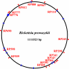
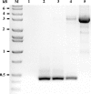
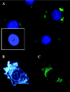
Rickettsia prowazekii, the causative agent of epidemic typhus, is an obligate intracellular bacterium that grows directly within the cytoplasm of its host cell, unbounded by a vacuolar membrane. The obligate intracytoplasmic nature of rickettsial growth places severe restrictions on the genetic analysis of this distinctive human pathogen. In order to expand the repertoire of genetic tools available for the study of this pathogen, we have employed the versatile mariner-based, Himar1 transposon system to generate insertional mutants of R. prowazekii. A transposon containing the R. prowazekii arr-2 rifampin resistance gene and a gene coding for a green fluorescent protein (GFP(UV)) was constructed and placed on a plasmid expressing the Himar1 transposase. Electroporation of this plasmid into R. prowazekii resulted in numerous transpositions into the rickettsial genome. Transposon insertion sites were identified by rescue cloning, followed by DNA sequencing. Random transpositions integrating at TA sites in both gene coding and intergenic regions were identified. Individual rickettsial clones were isolated by the limiting-dilution technique. Using both fixed and live-cell techniques, R. prowazekii transformants expressing GFP(UV) were easily visible by fluorescence microscopy. Thus, a mariner-based system provides an additional mechanism for generating rickettsial mutants that can be screened using GFP(UV) fluorescence.
Figures


Similar articles
-
Transposon mutagenesis of the obligate intracellular pathogen Rickettsia prowazekii.Appl Environ Microbiol. 2004 May;70(5):2816-22. doi: 10.1128/AEM.70.5.2816-2822.2004. Appl Environ Microbiol. 2004. PMID: 15128537 Free PMC article.
-
Transformation frequency of a mariner-based transposon in Rickettsia rickettsii.J Bacteriol. 2011 Sep;193(18):4993-5. doi: 10.1128/JB.05279-11. Epub 2011 Jul 15. J Bacteriol. 2011. PMID: 21764933 Free PMC article.
-
Transformation of Rickettsia prowazekii to rifampin resistance.J Bacteriol. 1998 Apr;180(8):2118-24. doi: 10.1128/JB.180.8.2118-2124.1998. J Bacteriol. 1998. PMID: 9555894 Free PMC article.
-
Dissecting the Rickettsia prowazekii genome: genetic and proteomic approaches.Ann N Y Acad Sci. 2005 Dec;1063:35-46. doi: 10.1196/annals.1355.005. Ann N Y Acad Sci. 2005. PMID: 16481488 Review.
-
Progress in rickettsial genome analysis from pioneering of Rickettsia prowazekii to the recent Rickettsia typhi.Ann N Y Acad Sci. 2005 Dec;1063:13-25. doi: 10.1196/annals.1355.003. Ann N Y Acad Sci. 2005. PMID: 16481486 Review.
Cited by
-
Cell-selective proteomics reveal novel effectors secreted by an obligate intracellular bacterial pathogen.Nat Commun. 2024 Jul 18;15(1):6073. doi: 10.1038/s41467-024-50493-9. Nat Commun. 2024. PMID: 39025857 Free PMC article.
-
Toxin synthesis by Clostridium difficile is regulated through quorum signaling.mBio. 2015 Feb 24;6(2):e02569. doi: 10.1128/mBio.02569-14. mBio. 2015. PMID: 25714717 Free PMC article.
-
A patatin-like phospholipase mediates Rickettsia parkeri escape from host membranes.Nat Commun. 2022 Jun 27;13(1):3656. doi: 10.1038/s41467-022-31351-y. Nat Commun. 2022. PMID: 35760786 Free PMC article.
-
Recent molecular insights into rickettsial pathogenesis and immunity.Future Microbiol. 2013 Oct;8(10):1265-88. doi: 10.2217/fmb.13.102. Future Microbiol. 2013. PMID: 24059918 Free PMC article. Review.
-
Analysis of Orientia tsutsugamushi promoter activity.Pathog Dis. 2021 Sep 23;79(7):ftab044. doi: 10.1093/femspd/ftab044. Pathog Dis. 2021. PMID: 34515306 Free PMC article.
References
-
- Andersson, S. G. E., A. Zomorodipour, J. O. Andersson, T. Sicheritz-Ponten, U. C. M. Alsmark, R. M. Podowdki, A. K. Naslund, A.-S. Eriksson, H. H. Winkler, and C. G. Kurland. 1998. The genome sequence of Rickettsia prowazekii and the origin of mitochondria. Nature 396:133-143. - PubMed
-
- Ausubel, F., R. Brent, R. E. Kingston, D. D. Moore, J. G. Seidman, J. A. Smith, and K. Struhl. 1997. Current protocols in molecular biology, vol. 1, 2, and 3. John Wiley & Sons, Inc., New York, NY.
-
- Bullock, W. O., J. M. Fernandez, and J. M. Short. 1987. XL1-Blue: a high efficiency plasmid transforming recA Escherichia coli strain with beta-galactosidase selection. BioTechniques 5:376-379.
Publication types
MeSH terms
Substances
Grants and funding
LinkOut - more resources
Full Text Sources

