. 2007 Oct;73(20):6612-9.
doi: 10.1128/AEM.00855-07. Epub 2007 Aug 24.

Immunoglobulins to surface-associated biofilm immunogens provide a novel means of visualization of methicillin-resistant Staphylococcus aureus biofilms

Affiliations

Immunoglobulins to surface-associated biofilm immunogens provide a novel means of visualization of methicillin-resistant Staphylococcus aureus biofilms

Rebecca A Brady et al. Appl Environ Microbiol. 2007 Oct.

Abstract

Antigens from the methicillin-resistant Staphylococcus aureus (MRSA) cell wall have been shown to be immunogenic in vivo and upregulated during biofilm growth. In this study, we created purified, recombinant forms of selected antigens and biofilm-upregulated, cell wall-associated proteins. These proteins were shown to cause a robust polyclonal immunoglobulin G (IgG) response when used to immunize rabbits. Antibodies against these recombinant proteins bound to the native forms of each protein as harvested from in vitro grown biofilms of MRSA, as determined both via Western blot analysis and immunofluorescence confocal microscopy. These IgGs could be utilized as imaging tools that localize to areas of specific protein production within a biofilm. This work illustrates that immunogenic, cell wall-associated, biofilm-upregulated proteins are promising for in vitro visualization of biofilm growth, architecture, and space-function relationships.


Figures

FIG. 1.
FIG. 1.
Purified recombinant proteins elicit a strong antibody response. (A) Purified recombinant proteins were run on an SDS-PAGE gel and probed with convalescent-phase serum from the biofilm infection model. (B) Purified recombinant proteins were run on an SDS-PAGE gel and probed with serum drawn from rabbits vaccinated with individual recombinant proteins. (C) Total protein from the cell wall fraction of an in vitro grown biofilm were run on an SDS-PAGE gel and probed with serum drawn from rabbits immunized with individual recombinant proteins. (D) Coomassie-stained SDS-PAGE gel with 5 μg of each purified recombinant protein resolved. Arrows point to bands corresponding to the molecular masses of lipase, SA0486, SA0037, SA0688, and glucosaminidase (Gluco), predicted to be 36, 27, 33, 31, and 27 kDa, respectively.
FIG. 2.
FIG. 2.
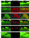
IgGs against recombinant forms of cell wall-associated biofilm proteins bind to intact MRSA biofilms. MRSA biofilms were grown as described previously (5), and IgG against each selected candidate protein was applied (A to E), followed by the secondary goat anti-rabbit F(ab′)2 antibody (red) (A to F). After a washing step, SYTO 9 was applied to stain all bacterial cells (green). Biofilms were probed with anti-lipase IgG and secondary antibody (A), anti-SA0486 IgG and secondary antibody (B), anti-SA0037 IgG and secondary antibody (D), anti-SA0688 IgG and secondary antibody (D), anti-glucosaminidase IgG and secondary antibody (E), and secondary antibody alone (as a negative control) (F). The base of the glass is located at the bottom of each image, and each image is a cross-sectional view of the biofilm from the base into the lumen. Size bar, 20 μm.
FIG. 3.
FIG. 3.
IgGs against some recombinant S. aureus proteins also bind to S. epidermidis biofilms. IgGs that show reactivity with the biofilm are seen in red. Total biofilm is shown in green. Goat anti-rabbit F(ab′)2 was used as a secondary antibody. Probes are as follows: anti-lipase IgG and secondary antibody (A), anti-SA0486 IgG and secondary antibody (B), anti-SA0037 IgG and secondary antibody (C), anti-SA0688 IgG and secondary antibody (D), anti-glucosaminidase IgG and secondary antibody (E), and secondary antibody only (F) (as a negative control). Size bar, 20 μm.

References

    1. Alam, M., M. Sultana, G. B. Nair, R. B. Sack, D. A. Sack, A. K. Siddique, A. Ali, A. Huq, and R. R. Colwell. 2006. Toxigenic Vibrio cholerae in the aquatic environment of Mathbaria, Bangladesh. Appl. Environ. Microbiol. 72:2849-2855. - PMC - PubMed
    1. Beenken, K. E., P. M. Dunman, F. McAleese, D. Macapagal, E. Murphy, S. J. Projan, J. S. Blevins, and M. S. Smeltzer. 2004. Global gene expression in Staphylococcus aureus biofilms. J. Bacteriol. 186:4665-4684. - PMC - PubMed
    1. Biswas, R., L. Voggu, U. K. Simon, P. Hentschel, G. Thumm, and F. Gotz. 2006. Activity of the major staphylococcal autolysin Atl. FEMS Microbiol. Lett. 259:260-268. - PubMed
    1. Bradford, M. M. 1976. A rapid and sensitive method for the quantitation of microgram quantities of protein utilizing the principle of protein-dye binding. Anal. Biochem. 72:248-254. - PubMed
    1. Brady, R. A., J. G. Leid, A. K. Camper, J. W. Costerton, and M. E. Shirtliff. 2006. Identification of Staphylococcus aureus proteins recognized by the mediated immune response to a biofilm infection. Infect. Immun. 74:3415-3426. - PMC - PubMed

Publication types

MeSH terms

LinkOut - more resources