

doi: 10.1261/rna.646007.
Epub 2007 Aug 24.
A novel monoclonal antibody against human Argonaute proteins reveals unexpected characteristics of miRNAs in human blood cells
Affiliations
- PMID: 17720879
- PMCID: PMC1986805
- DOI: 10.1261/rna.646007
Item in Clipboard
A novel monoclonal antibody against human Argonaute proteins reveals unexpected characteristics of miRNAs in human blood cells
RNA.
2007 Oct.
Abstract
Argonaute (Ago) proteins bind to microRNA (miRNAs) and short interfering RNAs (siRNAs) and form the core components of effector complexes that mediate miRNA and siRNA function. Currently, there is a paucity of reliable antibodies against mammalian Ago proteins, thus precluding studies of endogenous Ago proteins from tissues. Here we report the development of 2A8, a novel anti-Ago monoclonal antibody that recognizes human and mouse Ago proteins and efficiently immunoprecipitates miRNAs. We report the characterization of 2A8 and its use to clone miRNAs from human brain and from preparations of human polymorphonuclear leukocytes (neutrophils), which revealed a prevalent miRNA with unusual features.
Figures
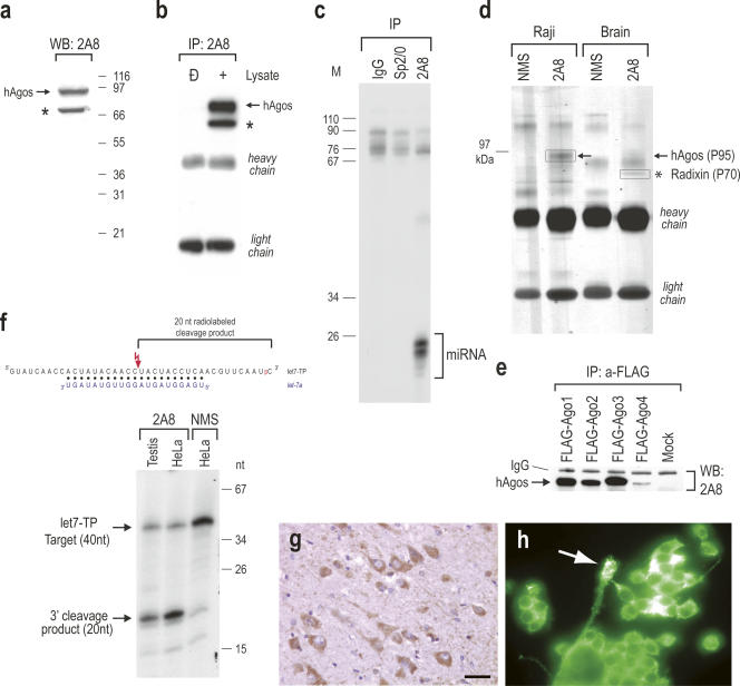
Properties of 2A8, a novel anti-Ago monoclonal antibody. (a) 2A8 recognizes Ago proteins on Western blot. Cell lysate from human HeLa cells was resolved on a 4%–12% NuPAGE gel, blotted and probed with 2A8. Molecular mass markers (in kilodaltons) are shown on the right. hAgos are shown with arrow. Asterisk indicates cross-reaction with radixin. (b) 2A8 immunoprecipitates Ago proteins. 2A8 immunoprecipitates from human 293 cells were resolved on a 4%–12% NuPAGE gel, blotted, and probed with 2A8 ascites. hAgos are shown with arrow. Asterisk indicates cross-reaction with radixin. (c) 2A8 coimmunoprecipitates miRNAs. Immunoprecipitations (IP) from HeLa cells were performed with 2A8 or nonimmune mouse serum (IgG) or Sp2/0 (both negative controls). RNA was isolated from the immunoprecipitates, 3′-end-labeled with [5′-32P]-pCp and T4 RNA ligase, resolved by electrophoresis on a 20% denaturing polyacrylamide gel, and visualized by autoradiography. Nucleotide sizes of the radiolabeled marker (M) are shown on the left. (d) Identification of immunoprecipitated protein with mass spectrometry. Immunoprecipitations were performed from human Raji cells or from human brain with 2A8 or nonimmune mouse serum (NMS). Immunoprecipitates were resolved on a 4%–12% NuPAGE gel, stained with Coomasie blue, and the P95 and P70 bands (boxed) were excised from the gel, subjected to trypsin digestion and mass spectrometry. (e) 2A8 recognizes all human Ago proteins. 293 cells were transfected with FLAG-tagged Ago1–4 proteins or with empty vector (Mock), followed by immunoprecipitations with anti-FLAG antibody. Immunoprecipitates were probed with 2A8 on Western blot. (f) 2A8 immunoprecipitates are active in miRNA-directed target RNA cleavage. Schematic of the RNA target (let7-TP) and the base-pairing with let-7a (blue); the [5′-32P] of pCp is shown in red. Cleavage site is indicated with red lightning bolt. Immunoprecipitations were performed with 2A8 or nonimmune mouse serum (NMS) and the beads were incubated with 3′-end-radiolabeled let7-TP RNA target. The products of the reactions were analyzed on 20% denaturing polyacrylamide gel. Nucleotide sizes of the radiolabeled marker (M) are shown on the right. (g,h) Immunocytochemistry with 2A8 was performed in formalin-fixed and paraffin-embedded (FFPE) human brain tissue (g) or MN-1, mouse motor neuron-like cells (h), and revealed predominantly cytoplasmic staining. In human FFPE brain, neurons were prominently stained (g). Arrow in h indicates a growth cone-like process of MN-1 cells, which is also labeled. The bar in (g) is either 50 μm (g) or 30 μm (h).
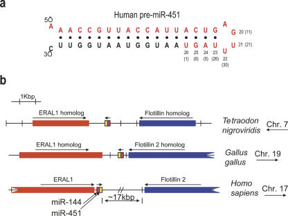
Cloning of miR-451 from immunopurified Agos from human neutrophils. (a) Sequence and secondary structure of human pre-miR-451. Mature miR-451 is shown in red. Nucleotide positions of the 3′-end of miR-451 are indicated. Percentages of clones that ended in specified nucleotide are shown in parentheses. Cloned miR-451 from human blood varied in length from 20 to 26 nt; 1% of clones were 26 nt long. (b) Genomic position of miR-451 gene in different species.
Similar articles
-
Expanded RNA-binding activities of mammalian Argonaute 2.Nucleic Acids Res. 2009 Dec;37(22):7533-45. doi: 10.1093/nar/gkp812. Nucleic Acids Res. 2009. PMID: 19808937 Free PMC article.
-
Anti-Argonaute RIP-Chip shows that miRNA transfections alter global patterns of mRNA recruitment to microribonucleoprotein complexes.RNA. 2010 Feb;16(2):394-404. doi: 10.1261/rna.1905910. Epub 2009 Dec 30. RNA. 2010. PMID: 20042474 Free PMC article.
-
Deep-sequencing of human Argonaute-associated small RNAs provides insight into miRNA sorting and reveals Argonaute association with RNA fragments of diverse origin.RNA Biol. 2011 Jan-Feb;8(1):158-77. doi: 10.4161/rna.8.1.14300. Epub 2011 Jan 1. RNA Biol. 2011. PMID: 21282978 Free PMC article.
-
MicroRNAs: biogenesis and molecular functions.Brain Pathol. 2008 Jan;18(1):113-21. doi: 10.1111/j.1750-3639.2007.00121.x. Brain Pathol. 2008. PMID: 18226106 Free PMC article. Review.
-
The Argonaute protein family.Genome Biol. 2008;9(2):210. doi: 10.1186/gb-2008-9-2-210. Epub 2008 Feb 26. Genome Biol. 2008. PMID: 18304383 Free PMC article. Review.
Cited by
-
A virus-derived microRNA targets immune response genes during SARS-CoV-2 infection.EMBO Rep. 2022 Feb 3;23(2):e54341. doi: 10.15252/embr.202154341. Epub 2021 Dec 16. EMBO Rep. 2022. PMID: 34914162 Free PMC article.
-
Antisense transcripts are targets for activating small RNAs.Nat Struct Mol Biol. 2008 Aug;15(8):842-8. doi: 10.1038/nsmb.1444. Epub 2008 Jul 6. Nat Struct Mol Biol. 2008. PMID: 18604220 Free PMC article.
-
Longer Work/Rest Intervals During High-Intensity Interval Training (HIIT) Lead to Elevated Levels of miR-222 and miR-29c.Front Physiol. 2018 Apr 17;9:395. doi: 10.3389/fphys.2018.00395. eCollection 2018. Front Physiol. 2018. PMID: 29719514 Free PMC article.
-
Consequences of depleting TNRC6, AGO, and DROSHA proteins on expression of microRNAs.RNA. 2023 Aug;29(8):1166-1184. doi: 10.1261/rna.079647.123. Epub 2023 May 11. RNA. 2023. PMID: 37169394 Free PMC article.
-
Expanded RNA-binding activities of mammalian Argonaute 2.Nucleic Acids Res. 2009 Dec;37(22):7533-45. doi: 10.1093/nar/gkp812. Nucleic Acids Res. 2009. PMID: 19808937 Free PMC article.
References
-
- Ambros, V. The functions of animal microRNAs. Nature. 2004;431:350–355. - PubMed
-
- Berezikov, E., Guryev, V., van de Belt, J., Wienholds, E., Plasterk, R.H., Cuppen, E. Phylogenetic shadowing and computational identification of human microRNA genes. Cell. 2005;120:21–24. - PubMed
-
- Bretscher, A. Regulation of cortical structure by the ezrin-radixin-moesin protein family. Curr. Opin. Cell Biol. 1999;11:109–116. - PubMed
-
- Cao, X., Yeo, G., Muotri, A.R., Kuwabara, T., Gage, F.H. Noncoding RNAs in the mammalian central nervous system. Annu. Rev. Neurosci. 2006;29:77–103. - PubMed
Publication types
MeSH terms
Substances
Grants and funding
LinkOut - more resources
Full Text Sources
Other Literature Sources