

Multi-disciplinary treatment of a rare pelvic cavity ependymoma
- PMID: 17722249
- PMCID: PMC2628065
- DOI: 10.3349/ymj.2007.48.4.719
Multi-disciplinary treatment of a rare pelvic cavity ependymoma
Abstract
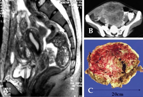
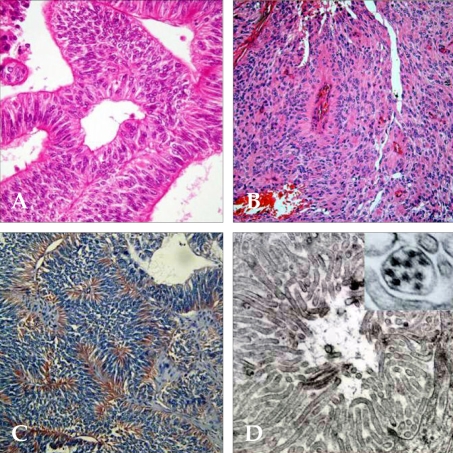
Ependymomas usually develop from neuroectodermal organs. Here, we present an ependymoma arising from the pelvic cavity. A 27-year-old Korean female was admitted to the hospital with a sensation of abdominal fullness. Imaging studies revealed a huge heterogeneous nodular mass in the pelvis and lower abdomen. Laparotomy showed that two large masses with multiple nodules were located between the uterus and rectum and uterus and bladder, respectively. Histologically, the tumor was characterized by compact columnar neoplastic cells divided by fibrovascular septae. The neoplastic cells formed true ependymal rosettes and perivascular pseudorosettes. Immunohistochemical staining showed a strong positive reaction for glial fibrillary acidic protein (GFAP) and vimentin and a partial positive reaction for S100 and EMA. The tumor was thus diagnosed as an ependymoma arising from the pelvic cavity. The patient was treated with a debulking operation and chemotherapy based upon the in vitro chemosensitivity test results. The patient was free of cancer for 4 years following surgery. This is a rare case of extraneural ependymoma for which an in vitro chemosensitivity test was critical in determining the multidisciplinary approach for treatment.
Figures
References
-
- Grody WW, Nieberg RK, Bhuta S. Ependymoma-like tumor of the mesovarium. Arch Pathol Lab Med. 1985;109:291–293. - PubMed
-
- Bell DA, Woodruff JM, Scully RE. Ependymoma of the broad ligament. A report of the two cases. Am J Surg Pathol. 1984;8:203–209. - PubMed
-
- Duggan MA, Hugh J, Nation JG, Robertson DI, Stuart GC. Ependymoma of the uterosacral ligament. Cancer. 1989;64:2565–2571. - PubMed
-
- Hofman V, Isnard V, Chevallier A, Motamedi JP, Michiels JF, Hassoun J, et al. Pelvic ependymoma arising from the small bowel. Pathology. 2001;33:26–29. - PubMed
-
- Dekmezian RH, Sneige N, Ordonez NG. Ovarian and omental ependymomas in peritoneal washings: cytologic and immunocytochemical features. Diagn Cytopathol. 1986;2:62–68. - PubMed
Publication types
MeSH terms
LinkOut - more resources
Full Text Sources
Research Materials
Miscellaneous
