

Micro- and nanofabrication methods in nanotechnological medical and pharmaceutical devices
- PMID: 17722281
- PMCID: PMC2676643
- DOI: 10.2147/nano.2006.1.4.483
Micro- and nanofabrication methods in nanotechnological medical and pharmaceutical devices
Abstract
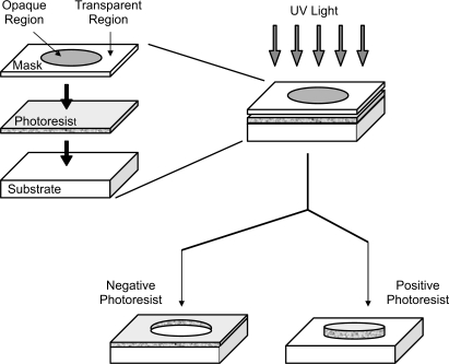
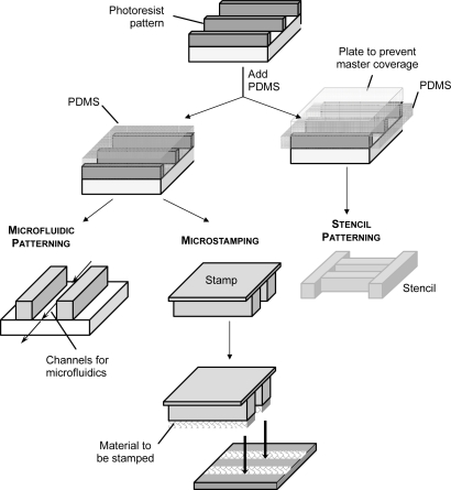
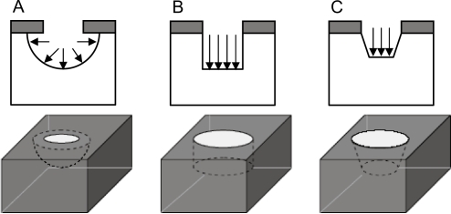
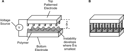
Micro- and nanofabrication techniques have revolutionized the pharmaceutical and medical fields as they offer the possibility for highly reproducible mass-fabrication of systems with complex geometries and functionalities, including novel drug delivery systems and bionsensors. The principal micro- and nanofabrication techniques are described, including photolithography, soft lithography, film deposition, etching, bonding, molecular self assembly, electrically induced nanopatterning, rapid prototyping, and electron, X-ray, colloidal monolayer, and focused ion beam lithography. Application of these techniques for the fabrication of drug delivery and biosensing systems including injectable, implantable, transdermal, and mucoadhesive devices is described.
Figures
Similar articles
-
Nanobiotechnology: protein-nanomaterial interactions.Biotechnol Prog. 2007 Mar-Apr;23(2):316-9. doi: 10.1021/bp060388n. Epub 2007 Mar 3. Biotechnol Prog. 2007. PMID: 17335286 Review.
-
Atomic layer deposition for biosensing applications.Biosens Bioelectron. 2018 Dec 30;122:147-159. doi: 10.1016/j.bios.2018.09.038. Epub 2018 Sep 13. Biosens Bioelectron. 2018. PMID: 30248642 Review.
-
Focused ion beam nanofabrication: a comparison with conventional processing techniques.J Nanosci Nanotechnol. 2006 Mar;6(3):661-8. doi: 10.1166/jnn.2006.111. J Nanosci Nanotechnol. 2006. PMID: 16573118 Review.
-
Nanofabrication of polymer surfaces utilizing colloidal lithography and ion etching.IEEE Trans Nanobioscience. 2006 Mar;5(1):9-14. doi: 10.1109/tnb.2005.864013. IEEE Trans Nanobioscience. 2006. PMID: 16570868
-
Fabrication of hierarchical micro-nanotopographies for cell attachment studies.Nanotechnology. 2013 Jun 28;24(25):255305. doi: 10.1088/0957-4484/24/25/255305. Epub 2013 May 31. Nanotechnology. 2013. PMID: 23727615
Cited by
-
Advancements in Micro/Nanorobots in Medicine: Design, Actuation, and Transformative Application.ACS Omega. 2025 Feb 4;10(6):5214-5250. doi: 10.1021/acsomega.4c09806. eCollection 2025 Feb 18. ACS Omega. 2025. PMID: 39989765 Free PMC article. Review.
-
An emerging era in manufacturing of drug delivery systems: Nanofabrication techniques.Heliyon. 2023 Mar 4;9(3):e14247. doi: 10.1016/j.heliyon.2023.e14247. eCollection 2023 Mar. Heliyon. 2023. PMID: 36938476 Free PMC article. Review.
-
A magneto-fluidic nanoparticle trapping platform for surface-enhanced Raman spectroscopy.Biomicrofluidics. 2017 Jun 7;11(3):034116. doi: 10.1063/1.4985071. eCollection 2017 May. Biomicrofluidics. 2017. PMID: 28652886 Free PMC article.
-
Geometry Control of Source/Drain Electrodes in Organic Field-Effect Transistors by Electrohydrodynamic Inkjet Printing.Materials (Basel). 2020 Nov 5;13(21):4974. doi: 10.3390/ma13214974. Materials (Basel). 2020. PMID: 33167331 Free PMC article.
-
Amperometric urea biosensors based on sulfonated graphene/polyaniline nanocomposite.Int J Nanomedicine. 2015 Aug 25;10 Spec Iss(Spec Iss):55-66. doi: 10.2147/IJN.S88315. eCollection 2015. Int J Nanomedicine. 2015. PMID: 26346240 Free PMC article.
References
-
- Andersson H, van den Berg A. Microfabrication and microfluidics for tissue engineering: state of the art and future opportunities. Lab on a Chip. 2004;4:98–103. - PubMed
-
- Bashir R, Hilt J, Elibol O, et al. Micromechanical cantilever as an ultrasensitive ph microsensor. Appl Phys Lett. 2002;81:3091–3.
-
- Becker H, Gärtner C. Polymer microfabrication methods for microfluidic analytical applications. Electrophoresis. 2000;21:12–26. - PubMed
-
- Blanchette JO, Kavimandan N, Peppas NA. Principles of transmucosal delivery of therapeutic agents. Biomed Pharmacother. 2004;58:142–51. - PubMed
-
- Bommannan D, Okuyama H, Stauffer P, et al. Sonophoresis. I. The use of high-frequency ultrasound to enhance transdermal drug delivery. Pharm Res. 1992;9:559–64. - PubMed
Publication types
MeSH terms
Substances
LinkOut - more resources
Full Text Sources
Other Literature Sources
