Review
. 2007;2(2):143-61.

Nanoparticulate carriers for the treatment of coronary restenosis

Affiliations
Review

Nanoparticulate carriers for the treatment of coronary restenosis

Luis Brito et al. Int J Nanomedicine. 2007.

Abstract

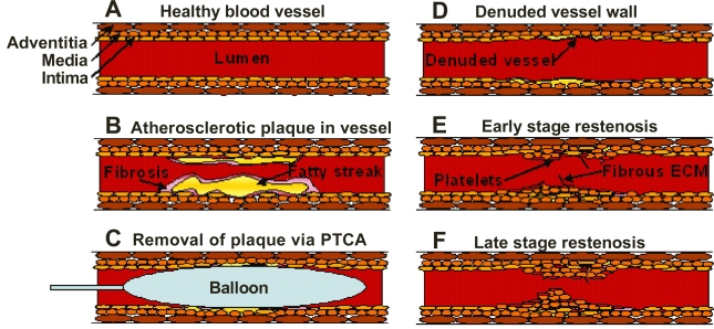
The current treatment for coronary restenosis following balloon angioplasty involves the use of a mechanical or a drug-eluting stent. Despite the high usage of commercially-available drug-eluting stents in the cardiac field, there are a number of limitations. This review will present the background ofrestenosis, go briefly into the molecular and cellular mechanisms of restenosis, the use of mechanical stents in coronary restenosis, and will provide an overview of the drugs and genes tested to treat restenosis. The primary focus of this article is to present a comprehensive overview on the use of nanoparticulate delivery systems in the treatment of restenosis both in-vitro and in-vivo. Nanocarriers have been tested in a variety of animal models and in human clinical trials with favorable results. Polymer-based nanoparticles, liposomes, and micelles will be discussed, in addition to the findings presented in the field of cardiovascular drug targeting. Nanocarrier-based delivery presents a viable alternative to the current stent based therapies.


Figures

Figure 1
Figure 1
Schematic illustration of the processes leading to restenotic lesion development. The figures show a healthy blood vessel (A), formation of atherosclerotic plaque within the blood vessel showing a fatty streak and macrophages encapsulated within a fibrotic tissue (B), insertion of a balloon angioplasty catheter to remove the plaque (C), damage due to stripping of the endothelial cells of the vessel wall after removal of the balloon (D), platelet accumulation and activation as well as rapid growth of smooth muscle cells and fibrous extracellular matrix forming the scaffolding (E), and the late stage restenosis showing neointima protruding into the lumen causing occlusion within the vessel (F).
Figure 2
Figure 2
The effect of time on arterial AG-1295 concentrations following poly(L-lactic acid) nanoparticle-based delivery. Drug levels are depicted on a logarithmic scale. Initial washout period occurs within 24 hours of administration and the drug levels can be seen for up to 14 days. The drug concentrations were measured by a high performance liquid chromatography assay. Insert shows confocal images of rat carotid arteries following local delivery of Nile Red® dye- containing fluorescent nanoparticles. Images were acquired at 5 minutes (A), 90 minutes (B), 1 day (C), 7 days (D) and 14 days (E), as well as, after 15 minutes of intraluminal delivery and 6 hours after delivery (F). Discrete, granular, fluorescent foci of nanoparticle aggregates are clearly observed in the image F. The notations L, M, N and A indicate lumen, media, neointima, and adventitia, respectively. Copyright © 2001. Reproduced from Fishbein I, Chorny M, Banai S, et al 2001. Formulation and delivery mode affect disposition and activity of tyrphostin-loaded nanoparticles in the rat carotid model. Arterioscler Thromb Vasc Biol, 21:1434–39.
Figure 3
Figure 3
Low magnification (10x) images of porcine arteries 4 weeks after stent implantation. The images represent stent only (A), untreated tissue as a negative control (B), and delivery of PCN1 ribozyme therapeutic agent (C). Panels A and B show well-defined intimal hyperplasia, whereas panel C shows a significantly lesser amount of neointimal growth. Lower panel images correspond to high magnification of the above. No histological evidence of tissue inflammation is seen. Copyright © 1999. Reproduced from Frimerman A, Welch PJ, Jin X, et al 1999. “Chimeric DNA-RNA hammerhead ribozyme to proliferating cell nuclear antigen reduces stent-induced stenosis in a porcine coronary model.” Circulation, 99:697–703.

Similar articles

Cited by

References

    1. Ahn YK, Kook H, Jeong MH, et al. Local RAD50 gene delivery induces regression of preformed porcine coronary in-stent neointimal hyperplasia. The Journal of Gene Medicine. 2004;6(1):93–104. - PubMed
    1. Armeanu S, Pelisek J, Krausz E, et al. Optimization of nonviral gene transfer of vascular smooth muscle cells in vitro and in vivo. Molecular Therapy. 2000;1(4):366–375. - PubMed
    1. Asahara T, Chen D, Tsurumi Y, et al. Accelerated restitution of endothelial integrity and endothelium-dependent function after phVEGF165 gene transfer. Circulation. 1996;94(12):3291–3302. - PubMed
    1. Axel DI, Kunert W, Goggelmann C, et al. Paclitaxel inhibits arterial smooth muscle cell proliferation and migration in vitro and in vivo using local drug delivery. Circulation. 1997;96(2):636–645. - PubMed
    1. Banai S, Chorny M, Gertz SD, et al. Locally delivered nanoencapsulated tyrphostin (AGL-2043) reduces neointima formation in balloon-injured rat carotid and stented porcine coronary arteries. Biomaterials. 2005;26(4):451–461. - PubMed

MeSH terms