

Sustained release of acyclovir from nano-liposomes and nano-niosomes: an in vitro study
- PMID: 17722549
- PMCID: PMC2673966
Sustained release of acyclovir from nano-liposomes and nano-niosomes: an in vitro study
Abstract
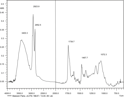
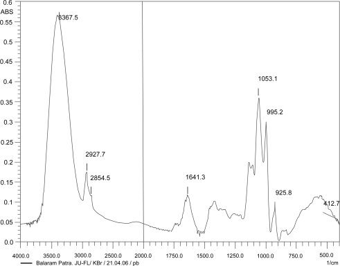
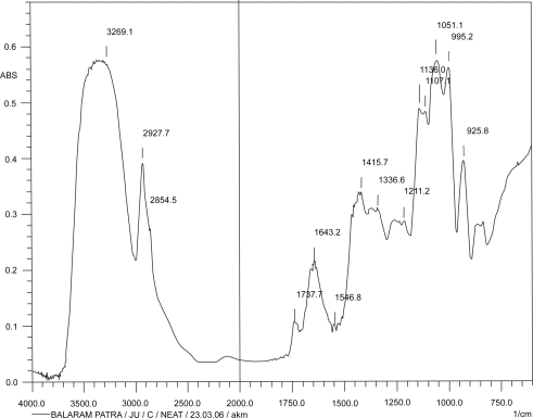
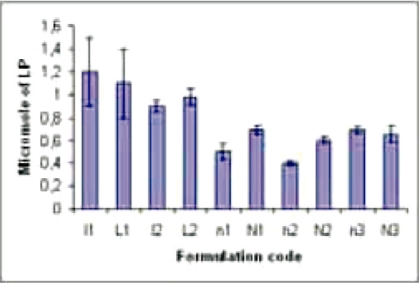
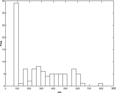
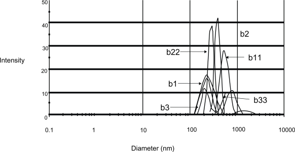
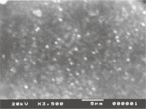
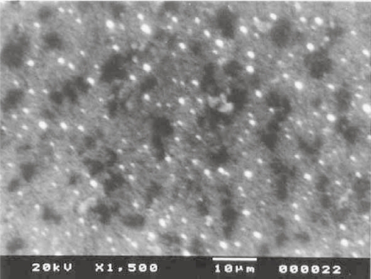
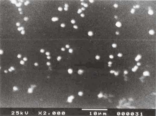
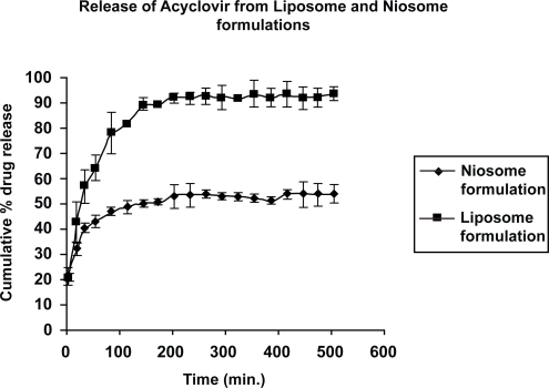
The present study was designed to develop and compare acyclovir containing nano-vesicular liposomes and niosomes based on cholesterol, soya L-alpha-lecithin and nonionic surfactant, span 20. The effort was made to study in vitro whether acyclovir-loaded nanovesicles could sustain the release of the drug by increasing residence time and thus, acyclovir could reduce its dose-related systemic toxicity. There were good vesicular distributions in both of the niosomes and the liposomes. The obtained vesicles were within 1 microm and about 35% of them were within a size of 100 nm. The percentage of drug loading varied and the niosomal vesicles contained more drug as compared with the liposomes. When the in vitro drug release was compared, it was found that the liposomes released about 90% drug in 150 min whereas the drug release was just 50% from the niosomal vesicles in 200 min. Again, the niosomes showed better stability compared with the liposomes. Thus, niosome could be a better choice for intravenous delivery of acyclovir.
Figures

References
-
- Brown RJ, Mc Crary M, Tyring SK. Antiviral agents, nonantiviral drugs. J Am Acad Dermatol. 2002;47:581–99. - PubMed
-
- Bundgared H, Jensen E, Falch E. Water soluble and solution stable and biolabile N-substituted (aminomethyl) benzoate ester prodrug of acyclovir. Pharm Res. 1991;8:1087–93. - PubMed
-
- Chetoni P, Rossi S, Burgalassi S, et al. Comparison of liposome-encapsulated acyclovir with acyclovir ointment: ocular pharmacokinetics in rabbits. J Ocul Pharmacol Ther. 2004;20:169–77. - PubMed
-
- Chikhale PJ, Venkatraghavan V, Bodor NS. Improved delivery through biological membranes: Intradermal targeting of acyclovir using redox based chemical drug delivery systems. Drug Del. 1996;3:17–26.
-
- Fang JY, Hong CT, Chiu WT, et al. Effect of liposomes and niosomes on skin permeation of enoxacin. Int J Pharm. 2001;219:61–72. - PubMed
Publication types
MeSH terms
Substances
LinkOut - more resources
Full Text Sources
