A freeze substitution fixation-based gold enlarging technique for EM studies of endocytosed Nanogold-labeled molecules
- PMID: 17723309
- PMCID: PMC2076746
- DOI: 10.1016/j.jsb.2007.07.004
A freeze substitution fixation-based gold enlarging technique for EM studies of endocytosed Nanogold-labeled molecules
Abstract
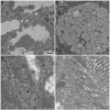
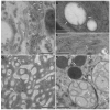
We have developed methods to locate individual ligands that can be used for electron microscopy studies of dynamic events during endocytosis and subsequent intracellular trafficking. The methods are based on enlargement of 1.4 nm Nanogold attached to an endocytosed ligand. Nanogold, a small label that does not induce misdirection of ligand-receptor complexes, is ideal for labeling ligands endocytosed by live cells, but is too small to be routinely located in cells by electron microscopy. Traditional pre-embedding enhancement protocols to enlarge Nanogold are not compatible with high pressure freezing/freeze substitution fixation (HPF/FSF), the most accurate method to preserve ultrastructure and dynamic events during trafficking. We have developed an improved enhancement procedure for chemically fixed samples that reduced auto-nucleation, and a new pre-embedding gold enlarging technique for HPF/FSF samples that preserved contrast and ultrastructure and can be used for high-resolution tomography. We evaluated our methods using labeled Fc as a ligand for the neonatal Fc receptor. Attachment of Nanogold to Fc did not interfere with receptor binding or uptake, and gold-labeled Fc could be specifically enlarged to allow identification in 2D projections and in tomograms. These methods should be broadly applicable to many endocytosis and transcytosis studies.
Figures


Similar articles
-
Silver enhancement of Nanogold particles during freeze substitution for electron microscopy.J Microsc. 2008 May;230(Pt 2):263-7. doi: 10.1111/j.1365-2818.2008.01983.x. J Microsc. 2008. PMID: 18445156 Free PMC article.
-
In situ localization of cartilage extracellular matrix components by immunoelectron microscopy after cryotechnical tissue processing.J Histochem Cytochem. 1987 Jun;35(6):647-55. doi: 10.1177/35.6.3553318. J Histochem Cytochem. 1987. PMID: 3553318
-
Nanogold as a specific marker for electron cryotomography.Microsc Microanal. 2009 Jun;15(3):183-8. doi: 10.1017/S1431927609090424. Microsc Microanal. 2009. PMID: 19460172 Free PMC article.
-
A practical technique to postfix nanogold-immunolabeled specimens with osmium and to embed them in Epon for electron microscopy.J Histochem Cytochem. 2000 Apr;48(4):493-8. doi: 10.1177/002215540004800407. J Histochem Cytochem. 2000. PMID: 10727291 Review.
-
Preparation and high-resolution microscopy of gold cluster labeled nucleic acid conjugates and nanodevices.Micron. 2011 Feb;42(2):163-74. doi: 10.1016/j.micron.2010.08.007. Epub 2010 Sep 8. Micron. 2011. PMID: 20869258 Free PMC article. Review.
Cited by
-
Silver enhancement of Nanogold particles during freeze substitution for electron microscopy.J Microsc. 2008 May;230(Pt 2):263-7. doi: 10.1111/j.1365-2818.2008.01983.x. J Microsc. 2008. PMID: 18445156 Free PMC article.
-
Effect of the First Feeding on Enterocytes of Newborn Rats.Int J Mol Sci. 2022 Nov 16;23(22):14179. doi: 10.3390/ijms232214179. Int J Mol Sci. 2022. PMID: 36430658 Free PMC article.
-
Synthesis, characterization, and direct intracellular imaging of ultrasmall and uniform glutathione-coated gold nanoparticles.Small. 2012 Jul 23;8(14):2277-86. doi: 10.1002/smll.201200071. Epub 2012 Apr 20. Small. 2012. PMID: 22517616 Free PMC article.
-
FcRn-mediated antibody transport across epithelial cells revealed by electron tomography.Nature. 2008 Sep 25;455(7212):542-6. doi: 10.1038/nature07255. Nature. 2008. PMID: 18818657 Free PMC article.
-
An intracellular traffic jam: Fc receptor-mediated transport of immunoglobulin G.Curr Opin Struct Biol. 2010 Apr;20(2):226-33. doi: 10.1016/j.sbi.2010.01.010. Epub 2010 Feb 18. Curr Opin Struct Biol. 2010. PMID: 20171874 Free PMC article. Review.
References
-
- Benlounes N, Chedid R, Thuillier F, Desjeux JF, Rousselet F, Heyman M. Intestinal transport and processing of immunoglobulin G in the neonatal and adult rat. Biol Neonate. 1995;67:254–263. - PubMed
-
- Berryman MA, Rodewald RD. An enhanced method for post-embedding immunocytochemical staining which preserves cell membranes. J Histochem Cytochem. 1990;38:159–170. - PubMed
-
- Burmeister WP, Gastinel LN, Simister NE, Blum ML, Bjorkman PJ. Crystal structure at 2.2 Å resolution of the MHC-related neonatal Fc receptor. Nature. 1994;372:336–343. - PubMed
-
- Busbee BD, Obare SO, Murphy CJ. An improved synthesis of high-aspect-ratio gold nanorods. Adv Mater. 2003;15:414–416.
-
- Daniel MC, Astruc D. Gold nanoparticles: Assembly, supramolecular chemistry, quantum-size-related properties, and applications toward biology, catalysis, and nanotechnology. Chemical Reviews. 2004;104:293–346. - PubMed
Publication types
MeSH terms
Substances
Grants and funding
LinkOut - more resources
Full Text Sources

