. 2007 Nov;75(11):5175-84.
doi: 10.1128/IAI.00719-07. Epub 2007 Aug 27.

Anthrax protective antigen cleavage and clearance from the blood of mice and rats

Affiliations

Anthrax protective antigen cleavage and clearance from the blood of mice and rats

Mahtab Moayeri et al. Infect Immun. 2007 Nov.

Abstract

Bacillus anthracis protective antigen (PA) is an 83-kDa (PA83) protein that is cleaved to the 63-kDa protein (PA63) as an essential step in binding and internalizing lethal factor (LF). To assess in vivo receptor saturating PA concentrations, we injected mice with PA variants and measured the PA remaining in the blood at various times using PA83- and PA63-specific enzyme-linked immunosorbent assays. We found that both wild-type PA (WT-PA) and a receptor-binding-defective mutant (Ub-PA) were cleaved to PA63 independent of their ability to bind cells. This suggested a PA-acting protease activity in the blood. The protease cleaved PA at the furin cleavage sequence because furin site-modified PA mutants were not cleaved. Cleavage measured in vitro was leupeptin sensitive and dependent on calcium. Cell surface cleavage was important for toxin clearance, however, as Ub-PA and uncleavable PA mutants were cleared at slower rates than WT-PA. The cell binding-independent cleavage of PA was also verified by using Ub-PA (which is still cleaved) to rescue mice from toxin challenge by competitively binding circulating LF. This mutant was able to rescue mice even when given 12 h before toxin challenge. Its therapeutic ability was comparable to that of dominant-negative PA, which binds cells but does not allow LF translocation, and to the protection afforded through receptor clearance by WT-PA and uncleavable receptor binding-competent mutants. The PA cleavage and clearance observed in mice did not appear to have a role in the differential mouse susceptibility as it occurred similarly in lethal toxin (LT)-resistant DBA/2J and LT-sensitive BALB/cJ mice. Interestingly, PA63 was not found in LT-resistant or -sensitive rats and PA83 clearance was slower in rats than in mice. Finally, to determine the minimum amount of PA required in circulation for LT toxicity in mice, we administered time-separated injections of PA and LF and showed that lethality of LF for mice after PA was no longer measurable in circulation, suggesting active PA sequestration at tissue surfaces.


Figures

FIG. 1.
FIG. 1.
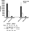
Monoclonal antibody detection of PA83 and PA63. (A) Detection of PA83 and PA63 by sandwich ELISA using 14B7 antibody for PA capture. Inset gel shows Western blot detection of PA83 and PA63 (2 ng/well loaded for each protein) probed with 14B7. (B) Similar ELISA experiment as in panel A, using monoclonal antibody 1G3. Inset gels show Western blots of PA83 and PA63 (8 ng/well loaded for each protein) with 1G3 antibody using Odyssey (LI-COR) blocking buffer or 5% skim milk. (C) Similar ELISA experiment as in panel A, using 1E9 PPT for PA capture. Inset gel shows concentrations detected by Western blotting of PA83 and PA63 with 1E9-PPT compared to anti-PA polyclonal antibody (4 ng/well loaded for each protein). ELISA results for panels A to C are representative of >50 ELISAs. (D) Detection of PA83 and PA63 by ELISA using 1E9-PPT, purified 1E9 ascites, and CCM1-PPT for antigen capture.
FIG. 2.
FIG. 2.
Clearance of PA from circulation. (A) BALB/cJ mice were injected i.v. with 5 to 400 μg of PA, and serum or plasma was collected 2 h later and analyzed for PA by ELISA using 14B7 monoclonal antibody as the capture antibody. The average values calculated from all experiments (serum and plasma collections) are shown numerically above each column. Standard deviation was calculated based on following n values with the indicated doses: 5 μg (n = 2 mice; six ELISAs), 10 μg (n = 4 mice; eight ELISAs), 20 μg (n = 6 mice; eight ELISAs), 50 μg (n = 4 mice; six ELISAs), 100 μg (n = 16 mice; eight ELISAs), 200 μg (n = 6 mice; six ELISAs), 400 μg (n = 10 mice; eight ELISAs). (B) PA concentrations in serum of BALB/cJ mice injected i.v. with 20, 100, or 200 μg of PA bled at 0.5, 2, 6, and 24 h were measured by 14B7 ELISA as follows: 0.5 h, n = 2 for all doses; 2 h, n = 6 for 20- and 200-μg doses and n = 16 for 100-μg dose; 6 h, n = 4 for 20- and 200-μg doses and n = 10 for 100-μg dose; and 24 h, n = 4 for all doses. All values are actual measured PA concentrations except zero time concentrations of PA, which were calculated based on an approximate 1.8-ml blood volume for the BALB/cJ mouse. All sigmoidal dose response (variable slope) analyses for calculations of concentrations based on PA ELISA standard curves and one-compartment half-life calculations were performed using GraphPad Prism, version 4.0, software. (C) Macrophage toxicity of serum collected at 2 h after injection of PA (50 μg, 200 μg, or 400 μg) in assays performed in vitro in the presence of 200 ng/ml or 20,000 ng/ml LF. Results are representative of four similar experiments performed with samples from two mice for each dose. (D) Comparison of PA concentrations in circulation at 2 h measured directly by ELISA compared to concentrations calculated from PA activity in the macrophage toxicity assay in the presence of LF in vitro. Standard deviations are based on averages from four experiments performed with duplicate samples from two individual mice per PA dose.
FIG. 3.
FIG. 3.
Cleavage and clearance of WT-PA and Ub-PA. (A) Concentrations of PA83 and PA63 detected in plasma collected at 2, 6, and 24 h after injection of PA (100 μg) measured by 14B7 and 1E9 ELISA (n = 16 mice per group). Western blot shows samples from two mice probed with anti-PA polyclonal (1:5,000). For Western blotting, plasma samples were diluted 50-fold (2 h) or 10-fold (6 h) prior to loading 5 μl/well. (B) Ub-PA83 and Ub-PA63 detected in plasma and serum collected at 2 h and 6 h after injection of Ub-PA (100 μg) by Western blots probed with anti-PA polyclonal (1:5,000). Samples were diluted 50-fold prior to loading 5 μl/well. (C) Comparison of PA83 plus PA63 measurements for WT-PA and Ub-PA in plasma at 2, 6, and 16 h after injection of 100 μg (i.v.) into BALB/cJ mice. The combined PA83 and PA63 measurements for samples from mice injected with WT-PA were assessed by 14B7 ELISA (n = 16 for 2 h and 6 h and n = 2 for 16 h). The combined PA83 and PA63 measurements for samples from mice injected with Ub-PA were assessed by 1G3 ELISA (n = 8 for 2 h and 6 h and n = 4 for 16 h) and are likely to be underestimates due to the low sensitivity of this ELISA compared to the 14B7 ELISA.
FIG. 4.
FIG. 4.
Cleavage and clearance of uncleavable PA mutants. (A) Western blots of three uncleavable PA mutants detected by polyclonal anti-PA (1:5,000) in plasma collected at various times after injection of 100 μg (i.v.) into BALB/cJ mice. Samples were diluted 50-fold (2 h) or 10-fold (6 h, 12 h, and 24 h) prior to loading 5 μl/well. Purified PA83 and PA63 controls are shown in first and last lanes. The PA63 oligomer is also seen in the last lane. (B) Western blots showing levels of the PA-U7 uncleavable mutant detected by polyclonal anti-PA (1:5,000) in plasma and serum collected at 2 h and 6 h after injection of 100 μg (i.v.) into BALB/cJ mice. Samples were diluted 50-fold prior to loading 3 μl/well. Purified PA83 and PA63 controls are in first, second, and last lanes. (C) Comparison of PA83 and PA63 concentrations as detected by 14B7 ELISA for WT-PA and three uncleavable PA mutants in plasma collected after injection of 100 μg into BALB/cJ mice. For WT-PA, n = 16 at 2 h, n = 16 at 6 h, n = 2 at 12 h, and n = 4 at 24 h, For PA-U7, n = 6 at 2 h, n = 6 at 6 h, n = 2 at 12 h, and n = 2 at 24 h. For PA-U2 and PA-L1, n = 4 at 2 and 6 h and n = 2 at 12 and 24 h.
FIG. 5.
FIG. 5.
Serum- and plasma-mediated cleavage of PA in vitro. (A) PA83 cleavage to PA63 in 15 mM HEPES-2 mM NaCl (4 left lanes) compared to DMEM-10 mM HEPES (four right lanes) using 1.5%, 6%, 12%, and 50% concentrations of BALB/cJ serum (S), EDTA-plasma (E), and heparin-plasma (H). Control lanes represent PBS treatment (P). (B) PA83 cleavage levels in 15 mM HEPES in the presence of CaCl2 (2 mM) and NaCl (100 mM) using 6% heparin-plasma (upper panel) or in DMEM with EDTA (50 mM)) using 6% heparin plasma and serum (lower panel). (C) Inhibition of PA83 cleavage to PA63 as quantified by PA63 ELISA using various protease inhibitors. Results shown are for the highest tested inhibitor doses listed in Materials and Methods and represent averages from two independent experiments. PPACK, d-phenylalanyl-l-arginine chloromethylketone. (D) Representative Western blots showing levels of inhibition of plasma-mediated PA83 cleavage (in DMEM) by leupeptin and aprotinin at various doses (shown in μg/ml).
FIG. 6.
FIG. 6.
Comparison of PA cleavage and clearance in BALB/cJ and DBA/2J mice. (A) Concentrations of PA83 and PA63 detected in plasma samples collected at 2 h and 6 h after injection of PA (100 μg) into BALB/cJ or DBA/2J mice measured by 14B7 and 1E9-PPT ELISAs (n = 4 mice per time group). Western blot shows samples from four mice probed with anti-PA polyclonal (1:5,000) antibody. Samples were diluted 50-fold (2 h) or 10-fold (6 h) prior loading 5 μl/well. PA63 control protein is shown in first well.
FIG. 7.
FIG. 7.
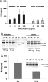
PA cleavage and clearance in rats. (A) Concentrations of PA83 detected by 14B7 ELISA in plasma samples collected at 2 and 6 h from Fischer rats injected with various doses of PA (n = 4 for each group). (B) Western blotting with plasma samples from Fischer or Lewis rats collected 2 or 6 h after injection of PA (100 μg or 5 μg). Western blot detection used anti-PA polyclonal antibody (1:5,000). (C) Concentration of PA83 detected by 14B7 ELISA and by Western blotting (anti-PA polyclonal antibody used at 1:5,000) in plasma samples collected at 6 and 18 h after injection of PA (100 μg) (n = 4 for each group in ELISAs).
FIG. 8.
FIG. 8.
PA clearance and LF toxicity in mice. (A) BALB/cJ mice were injected (i.v.) with various doses of PA (20, 50, 100, or 200 μg) followed by an immediate (0 h) or delayed (2, 4, 6, 8, 10, or 24 h) i.p. injection of LF alone (100 μg) and were monitored for survival (n = 4 mice per group). (B) BALB/cJ mice were injected (i.v.) with PA (100 μg), followed by an immediate (0 h) or delayed (6, 10, 16, or 24 h) i.p. injection of LF alone (100 μg) or LT (100 μg of PA plus 100 μg of LF) and were monitored for survival (n = 4 mice per group).
FIG. 9.
FIG. 9.
PA mutant protection against LT challenge. (A) BALB/cJ mice were injected i.v. (200 μl) with LT (60 μg of PA plus 20 μg of LF) alone or with Ub-PA included at various doses (60, 120, or 240 μg) and monitored for survival (n = 4 for each group). (B) BALB/cJ mice were injected i.v. (200 μl) with LT (35 μg of PA plus 15 μg of LF) alone or with Ub-PA included at 70 or 140 μg. In two groups of mice, Ub-PA (210 μg) was administered i.p. 30 min before or after the LT (35 μg of PA plus 15 μg of LF) injection (n = 4 for each group). (C) Groups of BALB/cJ mice were injected i.p. at 2, 6, or 12 h prior to LT challenge (i.v. injection of 35 μg of PA plus 15 μg of LF) with various doses of the following: PBS, WT-PA (WT), Ub-PA (Ub), PA-DN (DN), or U7-PA (U7). Mice were monitored for survival. Numbers in the table refer to the number of surviving mice relative to the number of mice in each treatment group.

Similar articles

Cited by

References

    1. Abrami, L., S. Liu, P. Cosson, S. H. Leppla, and F. G. van der Goot. 2003. Anthrax toxin triggers endocytosis of its receptor via a lipid raft-mediated clathrin-dependent process. J. Cell Biol. 160:321-328. - PMC - PubMed
    1. Bradley, K. A., J. Mogridge, M. Mourez, R. J. Collier, and J. A. Young. 2001. Identification of the cellular receptor for anthrax toxin. Nature 414:225-229. - PubMed
    1. Chekanov, A. V., A. G. Remacle, V. S. Golubkov, V. S. Akatov, S. Sikora, A. Y. Savinov, M. Fugere, R. Day, D. V. Rozanov, and A. Y. Strongin. 2005. Both PA63 and PA83 are endocytosed within an anthrax protective antigen mixed heptamer: A putative mechanism to overcome a furin deficiency. Arch. Biochem. Biophys. 446:52-59. - PubMed
    1. Duesbery, N. S., C. P. Webb, S. H. Leppla, V. M. Gordon, K. R. Klimpel, T. D. Copeland, N. G. Ahn, M. K. Oskarsson, K. Fukasawa, K. D. Paull, and G. F. Vande Woude. 1998. Proteolytic inactivation of MAP-kinase-kinase by anthrax lethal factor. Science 280:734-737. - PubMed
    1. Ezzell, J. W., Jr., and T. G. Abshire. 1992. Serum protease cleavage of Bacillus anthracis protective antigen. J. Gen. Microbiol. 138:543-549. - PubMed

Publication types

MeSH terms