. 2007 Nov;27(21):7497-510.
doi: 10.1128/MCB.00687-07. Epub 2007 Aug 27.

Conditional deletion of activating protein 2alpha (AP-2alpha) in the developing retina demonstrates non-cell-autonomous roles for AP-2alpha in optic cup development

Affiliations

Conditional deletion of activating protein 2alpha (AP-2alpha) in the developing retina demonstrates non-cell-autonomous roles for AP-2alpha in optic cup development

Erin A Bassett et al. Mol Cell Biol. 2007 Nov.

Abstract

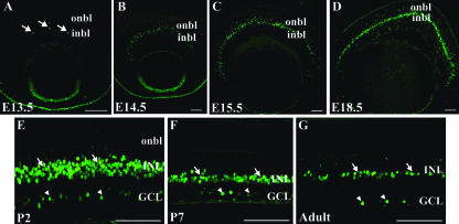
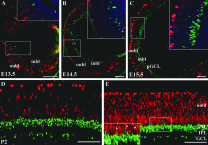
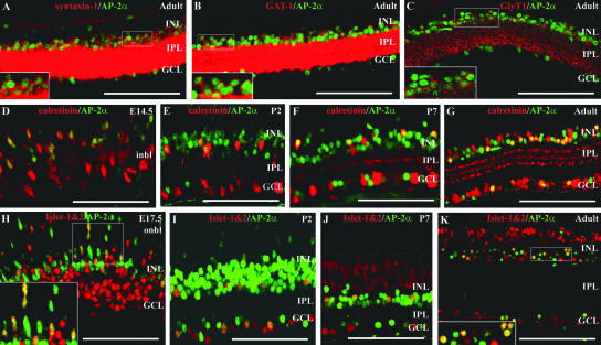
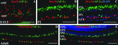
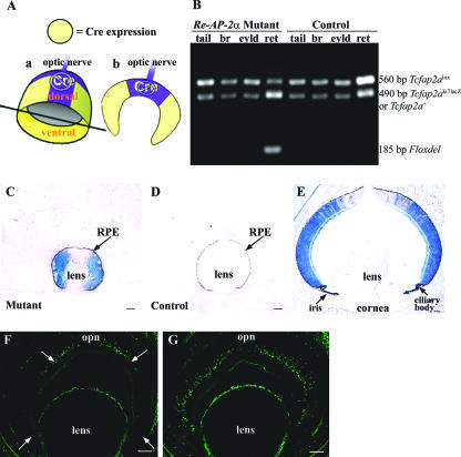
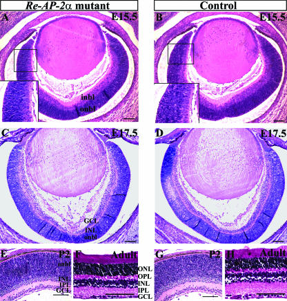
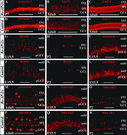
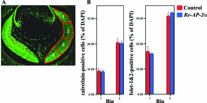
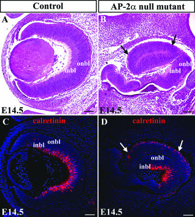
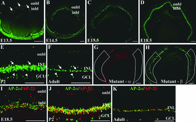
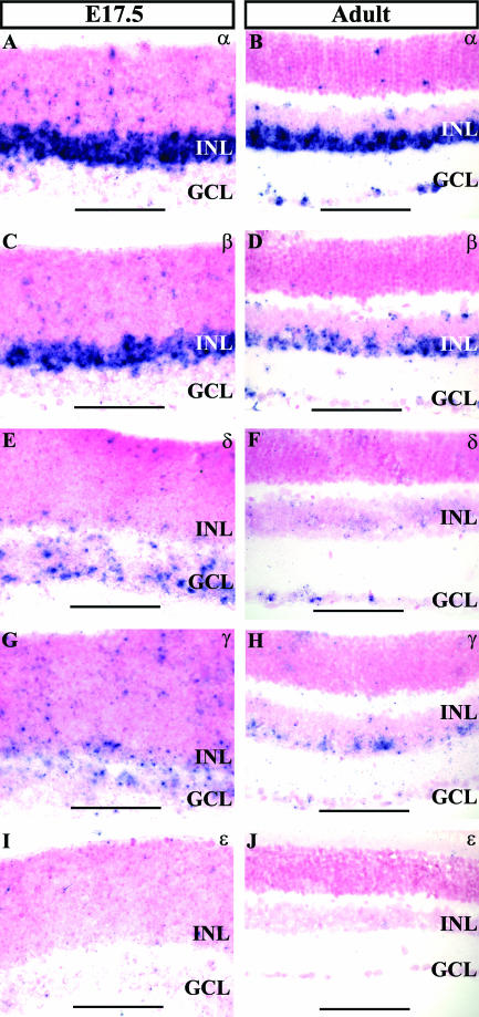
Activating protein 2alpha (AP-2alpha) is known to be expressed in the retina, and AP-2alpha-null mice exhibit defects in the developing optic cup, including patterning of the neural retina (NR) and a replacement of the dorsal retinal pigmented epithelium (RPE) with NR. In this study, we analyzed the temporal and spatial retinal expression patterns of AP-2alpha and created a conditional deletion of AP-2alpha in the developing retina. AP-2alpha exhibited a distinct expression pattern in the developing inner nuclear layer of the retina, and colocalization studies indicated that AP-2alpha was exclusively expressed in postmitotic amacrine cell populations. Targeted deletion of AP-2alpha in the developing retina did not result in observable retinal defects. Further examination of AP-2alpha-null mutants revealed that the severity of the RPE defect was variable and, although defects in retinal lamination occur at later embryonic stages, earlier stages showed normal lamination and expression of markers for amacrine and ganglion cells. Together, these data demonstrate that, whereas AP-2alpha alone does not play an intrinsic role in retinogenesis, it has non-cell-autonomous effects on optic cup development. Additional expression analyses showed that multiple AP-2 proteins are present in the developing retina, which will be important to future studies.


Figures

FIG. 1.
FIG. 1.
Expression pattern of the AP-2α protein in the developing mouse retina. Paraffin-embedded sections of embryos or dissected eyes were sectioned at 4 to 5 μm and stained with anti-AP-2α. (A) At E13.5, AP-2α initially appears in the central retina, in cells of the inner neuroblast layer (or presumptive INL; arrows). (B to D) As embryogenesis progresses, AP-2α expression extends to the periphery in a band of cells that is largely confined to the developing INL. (E to G) By P2 and continuing throughout adulthood, it is expressed in the INL (arrows), as well as a subset of cells in the GCL (arrowheads). i/onbl, inner/outer neuroblast layer. Scale bars, 100 μm.
FIG. 2.
FIG. 2.
AP-2α is not expressed in mitotic cells. Paraffin 4-μm horizontal sections from each developmental stage were double immunostained with anti-AP-2α (green) and the proliferation markers anti-phospho-histone H3 (A to D) or anti-Ki67 (E), both in red. No double-stained cells were detected in the retinas of embryonic (A to C) or neonatal (D and E) eyes. Squared areas are magnified at inset (top right; shown with nuclear stain DAPI [blue]). Arrowheads in panel E denote AP-2α-positive cells scattered in the highly proliferative outer neuroblast layer, which do not colocalize with Ki67. i/onbl, inner/outer neuroblast layer; pGCL, presumptive ganglion cell layer. Scale bars, 100 μm.
FIG. 3.
FIG. 3.
AP-2α colocalizes with syntaxin-1, GAT-1, GlyT1, calretinin, and Islet-1&2. Paraffin sections (4 to 5 μm) at different embryonic or postnatal stages were double stained with anti-AP-2α (green) and either anti-syntaxin-1, GlyT1, GAT-1, calretinin, or Islet-1&2 antibodies (red). A subset of AP-2α-positive cells colocalized with cells immunoreactive for syntaxin-1 (A), GAT-1 (B), GlyT1 (C), calretinin (D to G), and Islet-1&2 (H to K). Boxed areas in panels A to C, H, and K are magnified at the bottom left. i/onbl, inner/outer neuroblast layer. Scale bars, 100 μm.
FIG. 4.
FIG. 4.
AP-2α is not expressed in retinal ganglion cells. (A to C) Paraffin sections (4 μm) of E15.5 embryos and P2 eyes were double stained with anti-AP-2α (green) and anti-Brn3b (red). (B and C) Same section, shown with or without the nuclear stain DAPI (blue). (D to E) Fluorogold-labeled whole eyes were formalin fixed, paraffin embedded, and sectioned at 4 μm. The antigen retrieval necessary to detect AP-2α (shown in green) destroys Fluorogold fluorescence (shown in red); therefore, Fluorogold pictures were taken under the DAPI filter prior to immunostaining. (D and E) Same section, shown with or without DAPI-stained nuclei (blue). i/onbl, inner/outer neuroblast layer; ONL, outer nuclear layer; OPL, outer plexiform layer. Scale bars, 100 μm.
FIG. 5.
FIG. 5.
Cre recombinase activity and AP-2α deletion are specific to the peripheral retina. (A) Cre recombinase expression in α-Cre mice occurs in the peripheral aspects of the nasal and temporal NR but not in the central (i.e., toward the optic nerve head) and dorsal NR. Thus, cutting the retina horizontally at the optic nerve (in plane shown in subpanel a) results in sections (subpanel b) with Cre expression in the peripheral but not central retina. (B) Excision of Tcfap2a gene in mutant retina shown by PCR. Genomic DNA from the whole retina was examined by using primers Alflp, Alflox4, and Alfscsq (6) on Re-AP-2α mutants and control littermates. Tissues that do not express Cre recombinase display the 560-bp undeleted Tcfap2alox allele and a 490-bp product that represents either the Tcfap2aki7lacZ-null allele (in mutants) or a Tcfap2a+ (wild-type) copy (in littermates chosen as controls). The Tcfap2aki7lacZ and Tcfap2a+ alleles are distinguished in a separate PCR used for genotyping (not shown). In retinas of Re-AP-2α mutants, Cre recombinase acts on the loxP sites of the Tcfap2alox allele to excise the Tcfap2a gene, producing a shortened 185-bp “Floxdel” PCR product. Note that the Re-AP-2α mutants still contain a fraction of the unexcised Tcfap2alox allele due to the fact that Cre is not expressed throughout the entire retina. (C to E) α-Cre mice were bred with the ROSA26 Cre reporter strain (53), in which lacZ expression will occur upon removal of a floxed intervening segment by Cre recombinase. Horizontal sections of E13.5 (C and D) and P2 (E) heads were stained with X-Gal to detect lacZ activity, confirming the expected Cre activity in the peripheral retina. Black arrows denote the location of the RPE in E13.5 mutant and control. (F and G) Horizontal sections (at the optic nerve) of an E15.5 Re-AP-2α mutant (F) and a control littermate (G) stained with the anti-AP-2α antibody confirm regions lacking the AP-2α protein (white arrows) in the peripheral retina. br, brain; eyld, eyelid; ret, retina; opn, optic nerve. Scale bars, 100 μm.
FIG. 6.
FIG. 6.
Histology of Re-AP-2α and control retinas. H&E staining on 5-μm horizontal paraffin sections of E15.5 (A and B), E17.5 (C and D), P2 (E and G), and adult (F and H) Re-AP-2α mutants and control littermates. The sizes, shapes, and lamination of Re-AP-2α retinas resembled those of controls at all stages examined. Boxed areas in panels A and B are magnified in the insets (bottom left). i/onbl, inner/outer neuroblast layer; ONL, outer nuclear layer; OPL, outer plexiform layer. Scale bars, 100 μm.
FIG. 7.
FIG. 7.
Effect of retina-specific AP-2α deletion on the formation of different retinal cell types. Paraffin 4-μm horizontal sections that included the optic nerve were stained with either anti-syntaxin-1 (A and D), GlyT1 (B and E), GAT-1 (C and F), Brn3b (G, H, J, and K), calretinin (I, L, M, and P), or Islet-1&2 (N, O, Q, and R) antibodies. All pictures show the peripheral region, where excision of Tcfap2a occurs. Loss of AP-2α did not appear to result in a decrease in the number of cells immunoreactive for any markers tested. i/onbl, inner/outer neuroblast layer. Scale bars, 100 μm.
FIG. 8.
FIG. 8.
The number of calretinin- and Islet-1&2-positive cells is not altered in Re-AP-2α mutant retinas compared to control retinas. (A) An E15.5 horizontal section showing bins used for counting. The cells in bins 1 and 2 were quantified. (B) Quantification of cells immunoreactive for calretinin and Islet-1&2 at E15.5 in Re-AP-2α and control retinas. Counts are expressed as a percentage of DAPI-positive cells (shown as the mean ± the standard error for three retinas). The Student t test was used for statistical analysis. Loss of AP-2α did not alter the number of cells immunoreactive for calretinin or Islet-1&2 at E15.5.
FIG. 9.
FIG. 9.
Retinal lamination in AP-2α-null mutants proceeds normally during earlier stages of development. H&E stains of representative AP-2α-null mutant (B) and control littermate (A) eyes at E14.5. (B and D) The RPE defect (conversion of RPE to a second NR) is denoted by arrows. (C and D) Staining with anticalretinin showed that retinal lamination in the null mutants appeared to resemble that of control littermates. (D) The duplicated NR in AP-2α-null mutants expressed the inner NR marker calretinin on its outer surface. Scale bars, 100 μm.
FIG. 10.
FIG. 10.
Expression pattern of the AP-2β protein parallels that of AP-2α in the developing mouse retina. (A) At E13.5, AP-2β is expressed in the central retina, in cells of the inner neuroblast layer (or presumptive INL; arrows). (B to D) As embryogenesis progresses, AP-2β expression exhibits a central to peripheral expansion pattern in the developing INL. (E and F) By P2 and continuing throughout adulthood, AP-2β is expressed in the INL (arrows), as well as a subset of cells in the GCL (arrowheads). (G and H) Re-AP-2α mutant retinas immunostained for AP-2α (Mutant-α) or AP-2β (Mutant-β) illustrate that regions with AP-2α deletion (outlined in white) show strong expression of AP-2β. (I to K) AP-2β (red) colocalizes extensively with AP-2α (green) during embryonic (I) and postnatal (J) retinal development and in the adult retina (K). i/onbl, inner/outer neuroblast layer. Scale bars, 100 μm.
FIG. 11.
FIG. 11.
ISH analysis of all known AP-2 family members in the retina. (A to D) Transcripts of AP-2α and AP-2β are expressed in the developing INL at E17.5 and in the INL and GCL in the adult, in agreement with immunolocalization patterns. (E and F) AP-2δ mRNA predominates in the developing and mature GCL. (G and H) In the embryonic and adult retina, AP-2γ transcripts are expressed in the INL and GCL. (I and J) Signal for AP-2ɛ was not detected in the retina.

References

    1. Akagawa, K., M. Takada, H. Hayashi, and K. Uyemura. 1990. Calcium- and voltage-dependent potassium channel in the rat retinal amacrine cells identified in vitro using a cell type-specific monoclonal antibody. Brain Res. 518: 1-5. - PubMed
    1. Baumer, N., T. Marquardt, A. Stoykova, R. Ashery-Padan, K. Chowdhury, and P. Gruss. 2002. Pax6 is required for establishing naso-temporal and dorsal characteristics of the optic vesicle. Development 129: 4535-4545. - PubMed
    1. Bisgrove, D. A., and R. Godbout. 1999. Differential expression of AP-2α and AP-2β in the developing chick retina: repression of R-FABP promoter activity by AP-2. Dev. Dyn. 214: 195-206. - PubMed
    1. Bisgrove, D. A., E. A. Monckton, and R. Godbout. 1997. Involvement of AP-2 in regulation of the R-FABP gene in the developing chick retina. Mol. Cell. Biol. 17: 5935-5945. - PMC - PubMed
    1. Bosher, J. M., N. F. Totty, J. J. Hsuan, T. Williams, and H. C. Hurst. 1996. A family of AP-2 proteins regulates c-erbB-2 expression in mammary carcinoma. Oncogene 13: 1701-1707. - PubMed

Publication types

MeSH terms