. 2007 Aug 27;178(5):861-74.
doi: 10.1083/jcb.200705132.

Nectin-like proteins mediate axon Schwann cell interactions along the internode and are essential for myelination

Affiliations

Nectin-like proteins mediate axon Schwann cell interactions along the internode and are essential for myelination

Patrice Maurel et al. J Cell Biol. .

Abstract

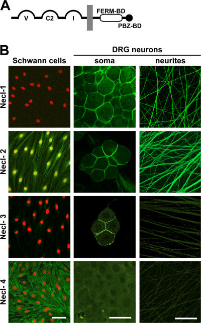
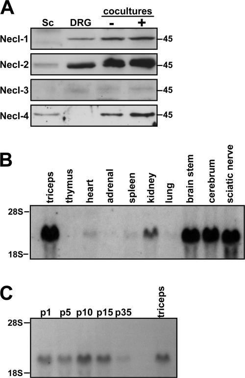
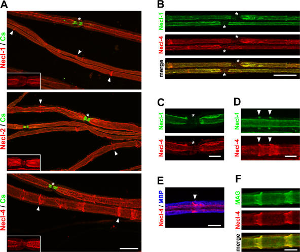
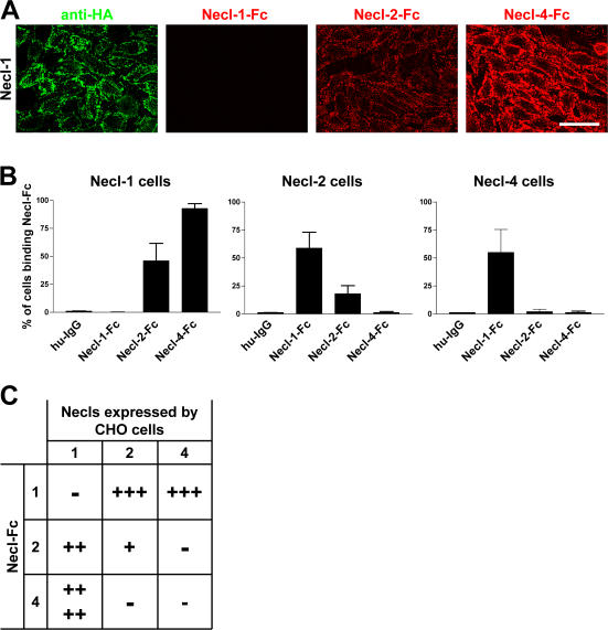
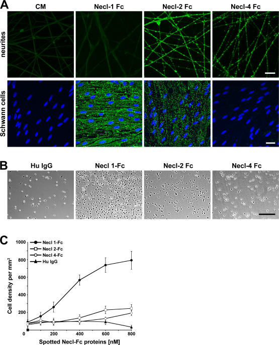
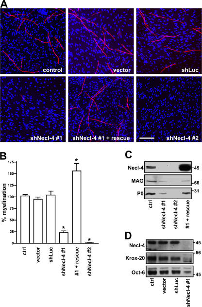
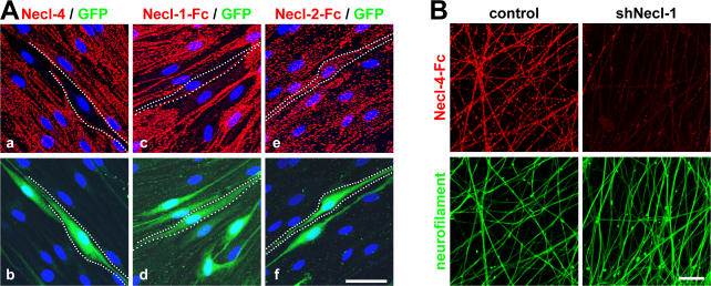
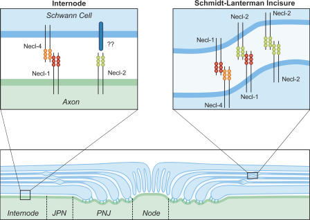
Axon-glial interactions are critical for the induction of myelination and the domain organization of myelinated fibers. Although molecular complexes that mediate these interactions in the nodal region are known, their counterparts along the internode are poorly defined. We report that neurons and Schwann cells express distinct sets of nectin-like (Necl) proteins: axons highly express Necl-1 and -2, whereas Schwann cells express Necl-4 and lower amounts of Necl-2. These proteins are strikingly localized to the internode, where Necl-1 and -2 on the axon are directly apposed by Necl-4 on the Schwann cell; all three proteins are also enriched at Schmidt-Lanterman incisures. Binding experiments demonstrate that the Necl proteins preferentially mediate heterophilic rather than homophilic interactions. In particular, Necl-1 on axons binds specifically to Necl-4 on Schwann cells. Knockdown of Necl-4 by short hairpin RNA inhibits Schwann cell differentiation and subsequent myelination in cocultures. These results demonstrate a key role for Necl-4 in initiating peripheral nervous system myelination and implicate the Necl proteins as mediators of axo-glial interactions along the internode.


Figures

Figure 1.
Figure 1.
Expression of Necl proteins by Schwann cells and DRG neurons. (A) Schematic organization of the Necl protein family. The extracellular segment contains three Ig domains of the variable (V), constant-2 (C2), and intermediate (I) types; the cytoplasmic region contains FERM- and PDZ-binding motifs as indicated. (B) Staining of cultured Schwann cells (left column) and of DRG somas and neurites (middle and right columns) with antibodies to Necl-1–4 is shown. Schwann cell nuclei are stained in red. Antibodies to Necl-1, -3, and -4 recognize the ectodomain of these proteins; the Necl-2 antibody reacts with its C terminus. Necl-1 specifically stains neurons, whereas Necl-4 specifically stains Schwann cells. Necl-2 stains Schwann cell membranes, nuclei, and neurons. Necl-1–3 accumulate at sites of contact between the neuronal cell somas. Bars, 50 μm.
Figure 2.
Figure 2.
Necl protein expression and Necl-4 mRNA expression. (A) Western blot analysis of Necl protein expression by cultured Schwann cells, DRG neurons, and cocultures under nonmyelinating (−) and myelinating (+) conditions. (B) Northern blot analysis of Necl-4 mRNA expression in various tissues. (C) Northern blot analysis of Necl-4 mRNA expression at different stages of sciatic nerve development.
Figure 3.
Figure 3.
The Necl proteins are localized in the internode and Schmidt-Lanterman incisures. (A) Teased sciatic nerves from adult mice were stained with antibodies to Necl-1, -2, or -4 (red) and Caspr (green), a marker of the paranodes. Antibodies to all three Necl proteins stain the internode and Schmidt-Lanterman incisures (arrowheads). Nodes, which are indicated by asterisks, are shown magnified in the insets without Caspr staining to demonstrate that Necl expression is largely excluded from the paranodes. (B) Teased sciatic nerves were double stained for Necl-1 (green) and -4 (red); the merged image is shown below. Two nodes and their flanking paranodes located in the center of the field are indicated with asterisks and are unlabeled. (C) A node of Ranvier stained for Necl-1 (green) and -4 (red). Both Necl-1 and -4 are largely excluded from the paranodes and node, which is indicated with asterisks. (D) An internodal segment of a myelinated nerve shows two incisures (arrowheads) that are stained with antibodies to Necl-1 (green) and -4 (red). In each case, Necl-1 expression is restricted to the outer portion of these clefts, whereas Necl-4 stains the entire cleft. (E) A segment of a myelinated nerve stained for Necl-4 (red) and MBP (blue) is shown. The arrowhead indicates a Schmidt-Lanterman incisure. (F) Necl-4 (red) colocalizes with MAG (green) along the glial internode and in the clefts as shown. Bars (A and B), 25 μm; (C–F) 10 μm.
Figure 4.
Figure 4.
Heterophilic and homophilic interactions of Necl proteins. (A) A representative experiment demonstrating the binding of Necl-Fc constructs to monolayers of Necl-1–transfected CHO cells. Staining for the HA epitope on Necl-1 (green) and of the bound Necl proteins (red) is shown. (B) Quantitation of the results of binding of individual Necl-Fcs to each Necl-expressing CHO cell line (mean ± SEM [error bars]). The most robust binding is between Necl-1 and -4. (C) Summary of binding in the Necl family. Binding is scored as no cells (−), <25% (+), 25–50% (++), 50–75% (+++), or 75–100% (++++) of cells with bound Necl-Fc. Bar, 50 μm.
Figure 5.
Figure 5.
Binding of Necl proteins to DRG neurons and Schwann cells. (A) Conditioned media from nontransfected cells (CM) or cells transfected with individual Necl-Fc constructs were incubated with cultures of DRG neurons (top) or Schwann cells (bottom). Bound Fc constructs were visualized with fluoresceinated anti–human IgG. Necl-1 Fc binds to Schwann cells but not neurons; Necl-4-Fc binds to neurons but not Schwann cells. (B) Representative phase images of Schwann cell binding to Necl-Fc proteins. Dissociated Schwann cells were incubated with plastic substrates spotted with human IgG (control) or purified Necl-Fc constructs. (C) Quantitation of the binding of Schwann cells to Necl-Fc constructs spotted onto dishes at the different concentrations indicated (mean ± SEM [error bars]). Schwann cells bound robustly only to the Necl-1–Fc substrate. Bars (A), 25 μm; (B) 200 μm.
Figure 6.
Figure 6.
Necl-4 expression is required for Schwann cell differentiation and myelination. (A) Representative images of myelinating cocultures in which control Schwann cells or Schwann cells infected with the lentiviral vector alone or lentiviruses encoding shRNA to a nonspecific sequence (shLuc) or to Necl-4 sequences, in one case rescued with an exogenous Necl-4 protein (shNecl-4 #1 + rescue), were added to DRG neurons under myelinating conditions. Myelin segments were stained for MBP (red) and with the Hoechst nuclear dye (blue). (B) Quantitation of the effects of Necl-4 knockdowns on Schwann cell myelination. Control Schwann cells or Schwann cells infected with the lentiviral vector alone or viruses encoding shRNAs to luciferase or Necl-4 with or without rescue by an shRNA-resistant Necl-4 were seeded onto dissociated DRG neuron cultures and maintained under myelinating conditions for 10 d. The number of myelin segments that formed were quantitated from three separate experiments (mean ± SEM [error bars]); controls were normalized to 100%. *, P < 0.001. (C) Knockdown of Necl-4 inhibits myelin protein expression. Lysates from control Schwann cells and cells infected with lentiviruses encoding shRNAs to Necl-4, including cells rescued with a modified Necl-4 protein, were blotted for Necl-4 and the myelin proteins MAG and P0; cultures correspond to those shown in Fig. 6 A. shRNA treatment effectively knocked down Necl-4 and myelin protein and was restored by the overexpression of an shRNA-resistant Necl-4. (D) Knockdown of Necl-4 inhibits the expression of myelin transcription factors. Western blot analysis demonstrates the substantial knockdown of Necl-4 and corresponding reductions of the transcription factors Krox-20 and Oct-6. Bar, 100 μm.
Figure 7.
Figure 7.
Necl-4 on Schwann cells binds to Necl-1 on axons. (A) Schwann cells were infected with a dilution of the lentivirus encoding shRNA (construct #1) to Necl-4. Effects of shRNA treatment on Necl-4 expression (a) and the binding of Necl-1–Fc (c) and Necl-2–Fc (e); corresponding fields shown below (b, d, and f) are stained for GFP to identify infected cells. Examples of infected cells are outlined by dashed white lines. (B) Necl-4–Fc does not bind to axons in the absence of Necl-1. shRNA-treated neurons were incubated with Necl-4–Fc; robust binding is seen in control but not shRNA-treated neurites. The corresponding Western blot is shown in Fig. S5 C (available at http://www.jcb.org/cgi/content/full/jcb.200705132/DC1). Bottom panels show the same fields stained for neurofilament. Bars (A), 25 μM; (B) 40 μm.
Figure 8.
Figure 8.
Summary of Necl protein interactions within domains of myelinated fibers. Schematic diagram showing the domain organization of a myelinated axon and the potential interactions between the Necl proteins in the internode and Schmidt-Lanterman incisures. In the internode and juxtaparanodes (JPN), Necl-1 on the axon binds to Necl-4 on the Schwann cell. Necl-2 is also present on the axon; its interacting partners on the Schwann cell are not yet known. The Necl proteins are also heavily expressed in the Schmidt-Lanterman incisures of mature myelinated nerves; Necl-1 and -2 are expressed primarily in the outer portions of the clefts (indicated), whereas Necl-4 is expressed in all layers. Potential interactions in the clefts of Necl-1 with Necl-4 and -2 and homophilic interactions of Necl-2 are illustrated. There is essentially no expression of the Necl proteins in the paranodal junctions (PNJ) or at the node. Necl proteins are shown as cis-dimers predicted from structural data.

Similar articles

Cited by

References

    1. Arase, N., A. Takeuchi, M. Unno, S. Hirano, T. Yokosuka, H. Arase, and T. Saito. 2005. Heterotypic interaction of CRTAM with Necl2 induces cell adhesion on activated NK cells and CD8+ T cells. Int. Immunol. 17:1227–1237. - PubMed
    1. Bateman, A., L. Coin, R. Durbin, R.D. Finn, V. Hollich, S. Griffiths-Jones, A. Khanna, M. Marshall, S. Moxon, E.L. Sonnhammer, et al. 2004. The Pfam protein families database. Nucleic Acids Res. 32:D138–D141. - PMC - PubMed
    1. Biederer, T. 2006. Bioinformatic characterization of the SynCAM family of immunoglobulin-like domain-containing adhesion molecules. Genomics. 87:139–150. - PubMed
    1. Biederer, T., Y. Sara, M. Mozhayeva, D. Atasoy, X. Liu, E.T. Kavalali, and T.C. Sudhof. 2002. SynCAM, a synaptic adhesion molecule that drives synapse assembly. Science. 297:1525–1531. - PubMed
    1. Boles, K.S., W. Barchet, T. Diacovo, M. Cella, and M. Colonna. 2005. The tumor suppressor TSLC1/NECL-2 triggers NK-cell and CD8+ T-cell responses through the cell-surface receptor CRTAM. Blood. 106:779–786. - PubMed

Publication types

MeSH terms