. 2007 Sep 3;204(9):2063-74.
doi: 10.1084/jem.20070868. Epub 2007 Aug 27.

MyD88-5 links mitochondria, microtubules, and JNK3 in neurons and regulates neuronal survival

Affiliations

MyD88-5 links mitochondria, microtubules, and JNK3 in neurons and regulates neuronal survival

Younghwa Kim et al. J Exp Med. .

Abstract

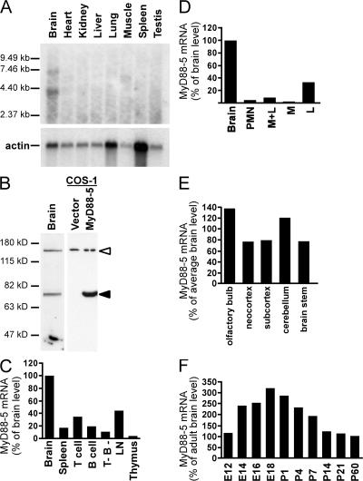
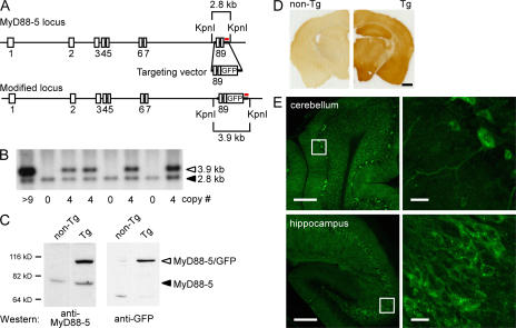
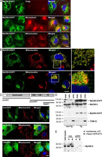
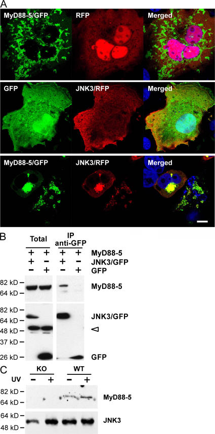
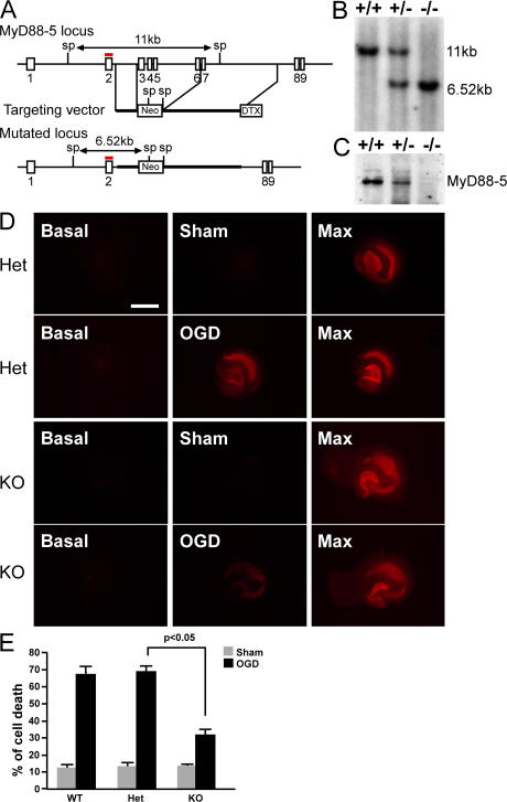
The innate immune system relies on evolutionally conserved Toll-like receptors (TLRs) to recognize diverse microbial molecular structures. Most TLRs depend on a family of adaptor proteins termed MyD88s to transduce their signals. Critical roles of MyD88-1-4 in host defense were demonstrated by defective immune responses in knockout mice. In contrast, the sites of expression and functions of vertebrate MyD88-5 have remained elusive. We show that MyD88-5 is distinct from other MyD88s in that MyD88-5 is preferentially expressed in neurons, colocalizes in part with mitochondria and JNK3, and regulates neuronal death. We prepared MyD88-5/GFP transgenic mice via a bacterial artificial chromosome to preserve its endogenous expression pattern. MyD88-5/GFP was detected chiefly in the brain, where it associated with punctate structures within neurons and copurified in part with mitochondria. In vitro, MyD88-5 co-immunoprecipitated with JNK3 and recruited JNK3 from cytosol to mitochondria. Hippocampal neurons from MyD88-5-deficient mice were protected from death after deprivation of oxygen and glucose. In contrast, MyD88-5-null macrophages behaved like wild-type cells in their response to microbial products. Thus, MyD88-5 appears unique among MyD88s in functioning to mediate stress-induced neuronal toxicity.


Figures

Figure 1.
Figure 1.
MyD88-5 is expressed mainly in brain, but not in myeloid cells. (A) Northern blot analysis of MyD88-5 expression in mouse tissues using the two SAM domains and part of the TIR domain (1,200–1,816 bp) as a probe. An actin probe was used as a loading control. (B) Western blot with chicken antibody raised against full-length recombinant mouse MyD88-5 reveals a 79-kD polypeptide in mouse brain (left lane) and in COS-1 cells transfected with a Flag-tagged MyD88-5 construct (right lane, filled arrowhead), but not in vector-transfected COS-1 cells (center lane). The open arrowhead indicates a nonspecific band. (C) Relative quantitation of MyD88-5 transcripts in mouse tissues and cells. Expression was highest in brain and moderate in lymph node (LN) and purified splenic T cells, but was barely detectable in splenic populations (chiefly macrophages) depleted of T and B cells (T- B-). Data are quantitative real-time PCR results normalized to mouse GAPDH and expressed as the percentage of the level in brain. One of three independent experiments is shown. (D) Relative quantitation of MyD88-5 transcripts in human tissues and cells. Expression of human MyD88-5 mRNA is highest in brain and detectable in lymphocytes (L). Polymorphonuclear leukocytes (PMN), monocytes (M), and lymphocytes were purified from human blood. Data are expressed as in C for one of three independent experiments. (E) MyD88-5 expression in various anatomic regions of adult mouse brain. Data are expressed as in C, and are from one of two independent experiments. (F) MyD88-5 expression in mouse brain during development. E12-18, embryonic days after conception; P1-60, postnatal days. qRT-PCR results are normalized to GAPDH and shown as the percentage of MyD88-5 expression in adult brain.
Figure 2.
Figure 2.
Generation of transgenic mice expressing MyD88-5-GFP fusion protein and its expression in neurons in the brain. (A) Schematic of the MyD88-5 locus in BAC clone RP23-399H5, the targeting vector, and the predicted structure of the modified MyD88-5 locus. Exon numbers are indicated. Probe used for Southern blotting is shown in red. Restriction sites and predicted fragment sizes are indicated. The stop codon in exon 9 is removed, and GFP is introduced in-frame. (B) Representative Southern blot of restricted genomic DNA from transgenic (Tg) and nontransgenic mice. Transgene copy numbers shown below are calculated by comparing the intensity of the endogenous band (filled arrowhead) and transgenic band (open arrowhead). (C) Expression of endogenous MyD88-5 and MyD88-5/GFP fusion protein in transgenic mouse brain. (left) Western blot with anti–MyD88-5 antibody. (right) Western blot with anti-GFP antibody. The fusion protein has the expected molecular weight of 105 kD. (D) Widespread expression of MyD88-5/GFP in the brain of transgenic mice. Brain sections from a nontransgenic mouse (left) and a transgenic (right) mouse were stained together with anti-GFP antibody. (E) Neuronal expression of MyD88-5/GFP. Fluorescent confocal microscopy captured direct GFP signals from unstained brain sections from a transgenic founder mouse. Right images are magnifications of the insets from the left. Bars: (D) 1 mm; (E, left) 200 μm; (E, right) 20 μm.
Figure 3.
Figure 3.
Mitochondrial association of MyD88-5. (A) Selective colocalization of MyD88-5/GFP fusion protein with mitochondria and microtubules. COS-1 cells were transfected with a MyD88-5/GFP fusion construct. 18 h after the transfection, cells were stained with MitoTracker Red CMXRos for 20 min and fixed, or fixed, permeabilized, and stained with antibody for Golgi (anti–γ-adaptin) or microtubules (anti-tubulin). (B) Extrinsic association of MyD88-5 with mitochondria. Confocal microscopic images of COS-1 cells transfected with MyD88-5/GFP and stained with MitoTracker Red CMXRos. (top) Cells expressing lower levels of MyD88-5; (bottom) cells expressing higher levels of MyD88-5, with effects on mitochondrial shape and localization. (right) Magnified images of the boxed areas from the images on the left. (C) Analysis of domains of MyD88-5 contributing to subcellular localization and mitochondrial association. COS-1 cells were transfected with the indicated constructs. 18 h later, mitochondria were stained with MitoTracker Red CMXRos before fixation. (D) Partial copurification of endogenous MyD88-5 and transgenic MyD88-5/GFP with mitochondria from brain. Mitochondria were isolated from wild type (non-Tg) or transgenic (Tg) mouse brains by differential centrifugation and lysates were Western blotted with antibody to MyD88-5 (top), GFP (middle), or TOM-23 (bottom). TOM-23 is an intrinsic mitochondrial membrane protein whose relative abundance serves as a loading control. (E) Copurification of endogenous MyD88-5 and microtubules from mouse brain. Microtubules were either polymerized with Taxol and GTP at 37°C or prevented from polymerization with colchicine at 4°C before purification by sucrose gradient centrifugation. Purified microtubule fractions (pellet), unpolymerized fractions (sup), and total brain lysate (total lysate) were Western blotted with antibody to MyD88-5. Bars, 10 μm.
Figure 4.
Figure 4.
MyD88-5–dependent recruitment of JNK3 to mitochondria. (A) Redistribution of JNK3 to mitochondria in the presence of MyD88-5. COS-1 cells were transfected with MyD88-5/GFP and RFP (top), GFP and JNK3/RFP (middle), or MyD88-5/GFP and JNK3/RFP (bottom). Mitochondria were stained with MitoTracker Red CMXRos. Bar, 10 μm. (B) Coimmunoprecipitation of MyD88-5 with JNK3/GFP, but not with GFP alone, from transfected COS-1 cells. Immunoprecipitation was with anti-GFP antibody and protein G–Sepharose and Western blotting was with antibody to MyD88-5 (top) or GFP (bottom). Open arrowhead indicates nonspecific bands. (C) Coimmunoprecipitation of MyD88-5 with JNK3 in hippocampal slices from wild-type (WT) or MyD88-5 knockout (KO) mice. Half of the brain slices were preexposed to 120 Joules/m2 of UV light. Immunoprecipitation was done with anti-JNK1,3 and protein G–Sepharose and Western blot with anti-JNK3 and anti-MyD88-5.
Figure 5.
Figure 5.
Generation of MyD88-5 knockout mice and protection of their hippocampal neurons from OGD-induced cell death. (A) Schematic of the MyD88-5 locus, targeting vector, and predicted structure of the mutated locus. Exon numbers are indicated. The probe used for Southern blotting is shown in red. Restriction enzyme sites and predicted sizes of the digests are shown. Sp, SphI; Neo, neomycin resistance gene; DTX, diphtheria toxin gene. (B) Southern blot analysis of genomic DNA from MyD88-5 wild-type, hemizygous-deficient, and null mice. (C) Western blot analysis of MyD88-5 expression in wild-type, hemizygous-deficient, and null mice. 50 μg of brain lysates were subjected to SDS-PAGE. Affinity-purified chicken anti–MyD88-5 antibody was used for detection. (D) Propidium iodide staining of dead neurons in hippocampal slices at the start of the OGD period (basal), after OGD (OGD), or the same period without OGD treatment (sham), and after exposure to 1 mM NMDA to induce a maximal cell death (Max). Hippocampi illustrated were from MyD88-5+/− (Het) or MyD88-5−/− mice (KO). Bar, 1 mm. (E) Quantitative evaluation of cell death after sham or OGD treatment in MyD88-5+/+ (sham, n = 6; OGD, n = 13) MyD88-5+/− (sham, n = 11; OGD, n = 22) and MyD88-5−/− (sham, n = 5; OGD, n = 29) slices. P < 0.05, Student's t test.

References

    1. Alexopoulou, L., A.C. Holt, R. Medzhitov, and R.A. Flavell. 2001. Recognition of double-stranded RNA and activation of NF-kappaB by Toll-like receptor 3. Nature. 413:732–738. - PubMed
    1. Xu, Y., X. Tao, B. Shen, T. Horng, R. Medzhitov, J.L. Manley, and L. Tong. 2000. Structural basis for signal transduction by the Toll/interleukin-1 receptor domains. Nature. 408:111–115. - PubMed
    1. Fitzgerald, K.A., E.M. Palsson-McDermott, A.G. Bowie, C.A. Jefferies, A.S. Mansell, G. Brady, E. Brint, A. Dunne, P. Gray, M.T. Harte, et al. 2001. Mal (MyD88-adapter-like) is required for Toll-like receptor-4 signal transduction. Nature. 413:78–83. - PubMed
    1. Horng, T., G.M. Barton, and R. Medzhitov. 2001. TIRAP: an adapter molecule in the Toll signaling pathway. Nat. Immunol. 2:835–841. - PubMed
    1. Yamamoto, M., S. Sato, H. Hemmi, H. Sanjo, S. Uematsu, T. Kaisho, K. Hoshino, O. Takeuchi, M. Kobayashi, T. Fujita, et al. 2002. Essential role for TIRAP in activation of the signalling cascade shared by TLR2 and TLR4. Nature. 420:324–329. - PubMed

Publication types

MeSH terms