. 2007 Sep;130(3):313-28.
doi: 10.1085/jgp.200709775.

Acute downregulation of ENaC by EGF involves the PY motif and putative ERK phosphorylation site

Affiliations

Acute downregulation of ENaC by EGF involves the PY motif and putative ERK phosphorylation site

Rebecca A Falin et al. J Gen Physiol. 2007 Sep.

Abstract

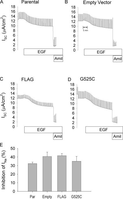
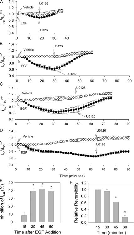
The epithelial sodium channel (ENaC) is expressed in a variety of tissues, including the renal collecting duct, where it constitutes the rate-limiting step for sodium reabsorption. Liddle's syndrome is caused by gain-of-function mutations in the beta and gamma subunits of ENaC, resulting in enhanced Na reabsorption and hypertension. Epidermal growth factor (EGF) causes acute inhibition of Na absorption in collecting duct principal cells via an extracellular signal-regulated kinase (ERK)-dependent mechanism. In experiments with primary cultures of collecting duct cells derived from a mouse model of Liddle's disease (beta-ENaC truncation), it was found that EGF inhibited short-circuit current (Isc) by 24 +/- 5% in wild-type cells but only by 6 +/- 3% in homozygous mutant cells. In order to elucidate the role of specific regions of the beta-ENaC C terminus, Madin-Darby canine kidney (MDCK) cell lines that express beta-ENaC with mutation of the PY motif (P616L), the ERK phosphorylation site (T613A), and C terminus truncation (R564stop) were created using the Phoenix retroviral system. All three mutants exhibited significant attenuation of the EGF-induced inhibition of sodium current. In MDCK cells with wild-type beta-ENaC, EGF-induced inhibition of Isc (<30 min) was fully reversed by exposure to an ERK kinase inhibitor and occurred with no change in ENaC surface expression, indicative of an effect on channel open probability (P(o)). At later times (>30 min), EGF-induced inhibition of Isc was not reversed by an ERK kinase inhibitor and was accompanied by a decrease in ENaC surface expression. Our results are consistent with an ERK-mediated decrease in ENaC open probability and enhanced retrieval of sodium channels from the apical membrane.


Figures

Figure 1.
Figure 1.
Effect of EGF on amiloride-sensitive ISC and ERK1/2 phosphorylation in primary renal collecting duct cells. (A) Confluent monolayers of primary renal collecting duct cells isolated from homozygous wild type (+/+) or Liddle's (L/L) mice were mounted in Ussing chambers and bathed on both sides with CT media. At the indicated time either EGF (20 ng/ml) or vehicle was added to the basolateral compartment. After 30 min, amiloride (100 μM) was added to the apical compartment. The vertical deflections are at 1-min intervals and represent the change in current when the voltage is clamped to a nonzero value to measure transepithelial resistance. (B) Time course for EGF-induced inhibition of amiloride-sensitive Isc (INa). Mean ± SEM for INa taken at 2-min intervals after addition of EGF (t = 0 min) to L/L (open circles; n = 8) or +/+ cells (closed circles; n = 9). (C) Summary of acute inhibition of INa 30 min after addition of EGF. n = 8–9. *, P < 0.05. (D) Western blot analysis of total and phosphorylated ERK1/2 in primary cultures of renal epithelial cells derived from either wild-type (+/+) or Liddle's (L/L) mice. Confluent monolayers were treated with EGF (20 ng/ml, basolateral) or vehicle for 30 min with and without a 15-min pretreatment with an ERK kinase inhibitor (U0126, 10 μM, apical/basolateral). Equivalent amounts of cell lysate (20 and 10 μg of cell lysate protein for total ERK1/2 and phosphorylated ERK1/2, respectively) were loaded into each lane. The blot is representative of three independent experiments.
Figure 2.
Figure 2.
Effect of EGF on amiloride-sensitive ISC in confluent monolayers of parental MDCK cells. Cells were seeded onto collagen-coated filters and grown in IM for 48 h. Filters were mounted in Ussing chambers and bathed on both sides with serum-free GM. (A) Representative trace. The epithelial monolayer was maintained under short-circuit conditions, and at 1-min intervals the voltage was clamped to a small nonzero value to measure transepithelial resistance. At the indicated time, EGF (20 ng/ml) was added to the basolateral compartment and 30 min later, amiloride (100 μM) was added to the apical compartment. (B and C) Summary of the effects of EGF and U0126 on INa and transepithelial resistance. Confluent monolayers of induced parental MDCK cells were mounted in Ussing chambers and after the current stabilized, vehicle (10 μl DMSO) or U0126 (10 μM, apical and basolateral) was added. 30 min later, vehicle or EGF (20 ng/ml, basolateral) was added followed 30 min later by amiloride (100 μM, apical). INa was calculated as the difference in ISC immediately before and after addition of amiloride. Transepithelial resistance was measured near the beginning of the experiment after the current stabilized. n = 5 for each group. *, P < 0.05.
Figure 3.
Figure 3.
Dose–response relationships for amiloride and benzamil inhibition of INa in engineered MDCK cell lines transduced with the FLAG-tagged β-ENaC. Validation of the G525C mutation. (A) Monolayers of MDCK cells stably expressing FLAG-tagged β-ENaC (circles; FLAG) or FLAG-tagged/G525C β-ENaC (triangles; G525C) were grown to confluence and mounted in Ussing chambers. Increasing concentrations of benzamil (blue) and amiloride (red) were added to the apical compartment and the relative INa determined as the ratio of the INa at the indicated concentration and that at the start of the experiment. (B) The fraction of total INa that was insensitive 1 μM benzamil (i.e., channels with a G525C β-ENaC) in MDCK cell lines stably expressing FLAG-tagged β-ENaC (FLAG) or FLAG-tagged/G525C β-ENaC (G525C).
Figure 4.
Figure 4.
Creation of the MDCK cell lines stably expressing engineered β-ENaC. (A) Diagrammatic representation of the modifications/mutations introduced into β-ENaC and stably expressed in MDCK cells. The name of each cell line is listed above and the modifications/mutations present within the channel are below the drawing. (B) Parental and clonal cell lines were harvested and analyzed for expression of the FLAG-tagged β-ENaC subunit. Anti-FLAG M2 antibody was used to immunoprecipitate FLAG-tagged β-ENaC from cell lysates. Bound proteins were eluted, separated by SDS-PAGE, and probed with an M2 anti-FLAG antibody. (C) Summary of INa in confluent monolayers of clonal MDCK cell lines stably expressing modified/mutated βENaC subunits (n = 5). INa was calculated as the fraction of ISC that was insensitive to 1 μM benzamil, but blocked by 10 mM amiloride. (D) Summary of transepithelial resistance in MDCK cell lines (n = 5).
Figure 5.
Figure 5.
Effect of EGF on short-circuit current in parental and modified MDCK cell lines. Parental MDCK cells were transduced with empty retroviral vector, FLAG-tagged wild-type β-ENaC, or FLAG-tagged/G525C β-ENaC (all transduced lines express GFP under the control of IRES). (A–D) Cells were grown to confluence on filters and mounted in Ussing chambers. After currents stabilized, EGF (20 ng/ml, basolateral) was added followed 30 min later by amiloride (10 mM, apical for G525C; 100 μM, apical for others). (E) Summary of EGF-induced inhibition of INa (n = 5 for each cell line).
Figure 6.
Figure 6.
Effect of EGF on short-circuit current in MDCK cell lines stably expressing mutant β-ENaC subunits. (A–D) Representative traces of short-circuit current in confluent monolayers of G525C, R564st, P616L, and T613A MDCK cell lines. At the indicated times benzamil (1 μM, apical), EGF (20 ng./ml, basolateral), and amiloride (10 mM, apical) were added. (E) The initial INa is the benzamil-insensitive, amiloride-sensitive, short-circuit current measured in three independent clonal cell lines for each mutation (n = 5 filters for each clone). (F) Summary of EGF-induced inhibition of INa in three independent clonal cell lines for each mutation (n = 5 filters for each clone). *, P < 0.05.
Figure 7.
Figure 7.
EGF-induced ERK1/2 phosphorylation and p-ERK1/2–dependent inhibition of INa in MDCK cell lines. G525C, R564st, P616, and T613A MDCK cell lines were grown to confluence on filters and bathed in IM for 48 h. (A) Individual monolayers were treated with vehicle (lane C; DMSO, 10 μl, both sides, 45 min), EGF (lane E; EGF, 20 ng/ml, basolateral, 15 min), ERK kinase inhibitor (U; U0126, 10 μM, both side, 45 min), or ERK kinase inhibitor plus EGF (U + E; U0126, 10 μM, both side, 30 then EGF, 20 ng/ml, basolateral, 15 min). Cells were then lysed, separated by SDS-PAGE, and probed for phosphorylated ERK1/2 and total ERK1/2. The blots are representative of three independent experiments. Densitometry was performed on each blot and the ratio of pERK1/2 to total ERK1/2 was determined for each cell line under each condition and the data are summarized in B. (C) Confluent monolayers of G525C, R564st, P616L, and T613A MDCK cell lines were exposed to EGF (20 ng/ml, 30 min, basolateral) with or without pretreatment with an ERK kinase inhibitor (U0126; 10 μM, 15 min, bilateral). The inhibition of INa was calculated as previously described. n = 5. P < 0.05. *, significantly different compared with G525C control; **, significantly different compared with monolayers not treated with U0126.
Figure 8.
Figure 8.
Time course for EGF-induced decrease in β-ENaC surface expression. (A) Surface expression of β-ENaC was determined by biotinylation and recovery of labeled apical membrane proteins by incubation with streptavidin beads. Biotinylated apical membrane and cytosolic protein samples were loaded on gels, separated by SDS-PAGE, transferred to nitrocellulose membranes, and probed with the M2 anti-FLAG antibody coupled to HRP to detect β-ENaC. The membranes were stripped and probed for actin. (B) Densitometry was performed and the ratio of surface to cytosol β-ENaC was determined for each sample and normalized to the ratio obtained at t = 0. n = 4. *, significantly different compared with ratio at t = 0. P < 0.05.
Figure 9.
Figure 9.
EGF-induced inhibition of INa in βG525C and βG525C/S518K mutant cell lines. Cells were grown to confluence on filters and mounted in Ussing chambers. After currents stabilized, benzamil (1 μM, apical) was added followed 5 min later by EGF (20 ng/ml, basolateral) addition, followed 30 min later by amiloride (10 mM, apical). (A) Initial INa was calculated as the ISC immediately before addition of EGF minus the Isc after addition of amiloride. (B) Summary of EGF-induced inhibition of INa (n = 6 for each cell line). *, P < 0.05.
Figure 10.
Figure 10.
Effect of EGF and β-ENaC mutations on activation of amiloride-sensitive short-circuit current by trypsin. (A) Representative trace of a parental MDCK cell line monolayer mounted in an Ussing chamber and exposed to apical trypsin (3 μg/ml) followed by amiloride (10 μM). (B) Representative trace of a parental MDCK cell line monolayer treated with apical amiloride (10 μM) followed by trypsin (3 μg/ml). (C) Confluent monolayers of G525C MDCK cells were mounted in Ussing chambers and treated with benzamil (1 μM, apical) to block endogenous sodium channels. 5 min later, either vehicle or EGF (20 ng/ml, basolateral) was added. At 35 min, trypsin (3 μg/ml, apical) was added, and at 50 min amiloride (10 mM, apical) was added. INa-trp/INa was calculated from the INa 15 min after trypsin addition divided by the INa immediately before trypsin addition. (D) G525C, P616L, and T613A MDCK cell line monolayers were mounted in Ussing chambers. Benzamil (1 μM, apical) was added at the start of the experiment, at 5 min trypsin (3 μg/ml, apical) was added followed by amiloride (10 mM, apical) at 20 min. The INa-trp/INa was calculated as above.
Figure 11.
Figure 11.
Reversibility of EGF-induced inhibition of amiloride-sensitive short-circuit current by an ERK kinase inhibitor. (A–D) Confluent monolayers of G525C MDCK cells were mounted in Ussing chambers and benzamil (1 μM) was added to the apical compartment. After the currents stabilized, EGF or vehicle (media) was added to the basolateral compartment. At various times thereafter (15, 30, 45, and 60 min), U0126 (10 μM) was added to the apical and basolateral compartments. Amiloride (10 mM) was added to the apical compartment at the end of experiment. The ISC t/ISC t=0 was calculated as the ratio of the ISC at each time point divided by the ISC at t = 0. Values represent mean ± SEM of the Isc recorded at 2-min intervals. (E) Summary of the time course for EGF-induced inhibition of amiloride-sensitive short-circuit current. EGF-induced inhibition in INa was calculated immediately before the addition of U0126 as difference between the ISC t /ISC t=0 for cells treated with EGF and those treated with vehicle. n = 5, P < 0.05. *, significantly different from zero. (F) The relative reversibility of EGF-induced inhibition of Isc was determined 30 min after the addition of U0126. It was calculated as the ISC t=end/ISC t=0 for EGF-treated cells divided by the ISC t=end/ISC t=0 for vehicle-treated cells. n = 5, P < 0.05. *, significantly different from 1.
Figure 12.
Figure 12.
Effect of ERK1/2 phosphorylation on INa following treatment with brefeldin A. (A and B) G525C and T613A MDCK cells grown to confluence were mounted in Ussing chambers, treated with benzamil (1 μM) to block channels with endogenous β-ENaC subunits and then exposed to vehicle or brefeldin A (20 μM; 30 min, bilateral) to inhibit delivery of new channels to the plasma membrane. At t = −5min, monolayers were treated with EGF (20 ng/ml, basolateral) or U0126 (10 μM, bilateral), and 30 min later amiloride (10 mM, apical) was applied. The relative INa was calculated at 1-min intervals as the INa at each time point relative to the INa at t = 0. n = 5 for each group.

Similar articles

Cited by

References

    1. Arnason, T., and M.J. Ellison. 1994. Stress resistance in Saccharomyces cerevisiae is strongly correlated with assembly of a novel type of multiubiquitin chain. Mol. Cell. Biol. 14:7876–7883. - PMC - PubMed
    1. Auberson, M., N. Hoffmann-Pochon, A. Vandewalle, S. Kellenberger, and L. Schild. 2003. Epithelial Na+ channel mutants causing Liddle's syndrome retain ability to respond to aldosterone and vasopressin. Am. J. Physiol. Renal Physiol. 285:F459–F471. - PubMed
    1. Blazer-Yost, B.L., X. Liu, and S.I. Helman. 1998. Hormonal regulation of ENaCs: insulin and aldosterone. Am. J. Physiol. 274:C1373–C1379. - PubMed
    1. Booth, R.E., and J.D. Stockand. 2003. Targeted degradation of ENaC in response to PKC activation of the ERK1/2 cascade. Am. J. Physiol. Renal Physiol. 284:F938–F947. - PubMed
    1. Bruns, J.B., M.D. Carattino, S. Sheng, A.B. Maarouf, O.A. Weisz, J.M. Pilewski, R.P. Hughey, T.R. Kleyman. 2007. Epithelial Na+ channels are fully activated by furin- and prostasin-dependent release of an inhibitory peptide from the γ subunit. J. Biol. Chem. 282:6153–6160. - PubMed

Publication types

MeSH terms