. 2007 Sep;48(9):3905-13.
doi: 10.1167/iovs.06-1464.

Analysis of ocular hypopigmentation in Rab38cht/cht mice

Affiliations

Analysis of ocular hypopigmentation in Rab38cht/cht mice

Brian P Brooks et al. Invest Ophthalmol Vis Sci. 2007 Sep.

Abstract

Purpose: To characterize the ocular phenotype resulting from mutation of Rab38, a candidate gene for Hermansky-Pudlak syndrome.

Methods: Chocolate mice (cht, Rab38(cht/cht)) and control heterozygous (Rab38(cht/)(+)) and wild-type mice were examined clinically, histologically, ultrastructurally, and electrophysiologically. Mice homozygous for both the Rab38(cht) and the Tyrp1(b) alleles were similarly examined.

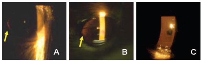
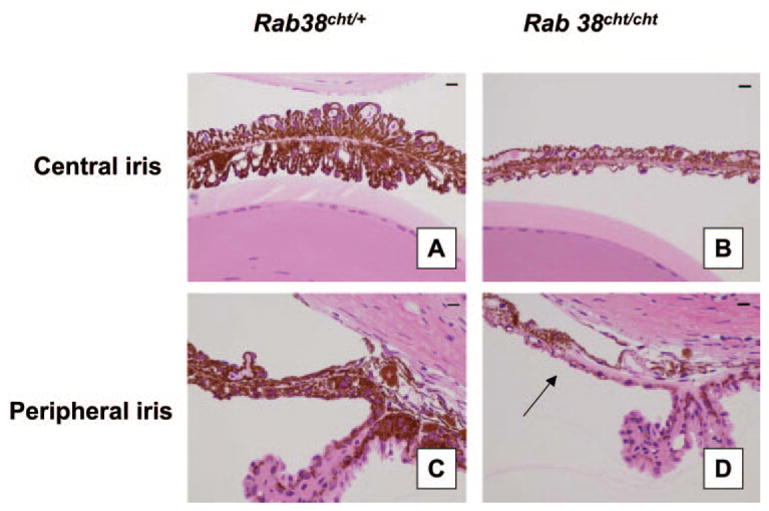
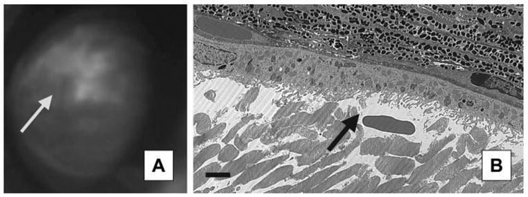
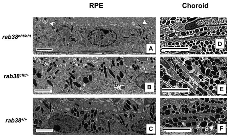
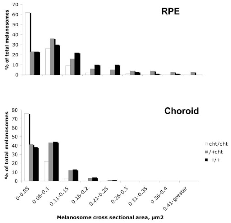
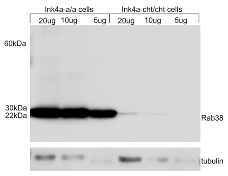
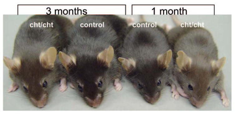
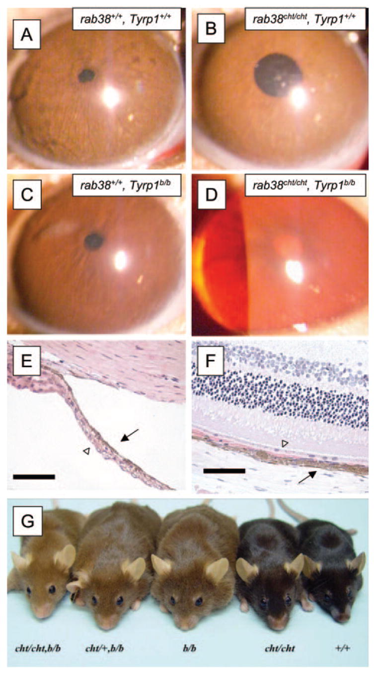
Results: Rab38(cht/cht) mice showed variable peripheral iris transillumination defects at 2 months of age. Patches of RPE hypopigmentation were noted clinically in 57% of Rab38(cht/cht) eyes and 6% of Rab38(cht/)(+) eyes. Rab38(cht/cht) mice exhibited thinning of the iris and RPE and larger b-wave amplitudes in the scotopic range when compared with the control animals. Compared with wild-type mice, Rab38(cht/cht) melanosomes were smaller and there were fewer in neuroectodermally derived retinal pigment epithelium; in neural crest-derived choroid melanocytes, they were smaller in size only. Mutation of both Rab38 and Tyrp1 produced mice with ocular and coat color pigment dilution greater than that seen with either mutation alone. Comprehensive clinical and pathologic analyses showed no other organ system or blood defects in Rab38(cht/cht) mice.

Conclusions: Rab38(cht/cht) mice show ocular characteristics reminiscent of human oculocutaneous albinism, as well as iris and RPE thinning. The synergistic effects of the Rab38(cht) and Tyrp1(b) alleles suggest that TYRP1 is not the only target of RAB38 trafficking. This mouse line provides a useful model for studying melanosome biology and its role in human ocular diseases.


Figures

Figure 1
Figure 1
Rab38cht/cht mice, but not Rab38cht/+ mice, exhibited peripheral iris transillumination (TI). The degree of TI was graded as +1 (peripheral only, A), +2 (peripheral and mid-iris, B) or 0 (none, C). Slightly more than 20% of young (2–6 months of age) Rab38cht/cht mice exhibited +2 TI; the remainder exhibited +1 TI. This proportion did not vary in a cohort of older mice (12–24 months), indicating that TI is probably due to variable iris hypoplasia, rather than iris atrophy. Rab38cht/+mice showed no iris TI, even at advanced ages (C, age 23 months in this instance). Similarly, wild-type mice did not exhibit iris TI (data not shown).
Figure 2
Figure 2
Histopathology of Rab38cht/cht mice (B, D) revealed iris hypopigmentation and thinning, particularly peripherally (D, arrow) when compared to that in age-matched Rab38cht/+ mice (A, C). Wild-type irides were similar to those of Rab38cht/+ mice (data not shown). Bars, 10 μm.
Figure 3
Figure 3
Rab38cht/cht mice exhibited peripheral patches of depigmentation (A, fundus photo). These patches were most frequently found in the superior retina. Transmission electron microscopy showed that they represented focal RPE thinning (B, arrow, border of normal-abnormal tissue dissected). Bar, 2 μm.
Figure 4
Figure 4
Melanosomes were smaller in cross-sectional area and fewer in the RPE of Rab38cht/cht mice (A) compared with wild-type (Rab38+/+; C); heterozygote (Rab38cht/+) mice were similar to wild-type (B). Melanosomes in Rab38cht/cht mice were able to enter the apical villi of RPE cells (A, arrowheads), suggesting that, unlike RAB27a, RAB38 is not necessary for transport into these structures. Melanosomes from the NC-derived choroid were also smaller in Rab38cht/cht mice than in wild-type mice. Bar, 2 μm.
Figure 5
Figure 5
The distribution of melanosome cross-sectional area in the RPE and choroid of Rab38cht/cht mice demonstrates the significantly smaller areas of Rab38cht/cht melanosomes relative to wild-type.
Figure 6
Figure 6
An immortalized pigmented mouse melanocyte cell line melan-Melan-Ink4a-cht5 had a reduced amount of RAB38 protein compared with the normal black pigmented cell line, melan-Ink4a-1, on Western blot.
Figure 7
Figure 7
Scotopic (A) and photopic (B) ERG b-wave amplitudes (±SEM from 2.5-month-old homozygous (Rab38cht/cht), heterozygous (Rab38cht/+), and wild-type C57BL/6J (Rab38+/+) mice. Compared with wild-type mice, homozygous mice had significantly larger b-wave amplitudes in the scotopic range (ANOVA, P < 0.01). In the photopic range, there was no significant difference between any of the groups. Homozygous and wild-type: n = 5 animals/10 eyes; heterozygous: n = 2 animals/4 eyes.
Figure 8
Figure 8
In one-month-old Rab38cht/cht mice, the light brown coat color was easily distinguished from the black coat of wild-type and Rab38cht/+ littermates. In 3-month-old and older Rab38cht/cht mice, the darker coat was still distinguishable from the black coat of the wild-type and Rab38cht/+ littermates.
Figure 9
Figure 9
The Tyrp1b/b allele interacted synergistically with the Rab38cht/cht allele in C57BI/6 mice, to produce striking pigment dilution. Slit lamp examination of the anterior segments of (A)Rab38+/+, Tyrp1+/+; (B) Rab38cht/cht, Tyrp1+/+; (C) Rab38+/+, Tyrp1b/b; and (D) Rab38cht/cht, Tyrp1b/b mice showed significantly more depigmentation and iris transillumination in the double-mutant homozygote than in the other three strains. This effect was observed in both the eyes and the coat color of the mice (G). (E, F) Rab38cht/cht, Tyrp1b/b mice showed reduced pigmentation on histologic sections, particularly in the neuroectodermally derived layers. This difference in pigmentation was evident in the posterior pigmented epithelial layer of the iris (E, arrowhead) and the RPE (F, arrowhead). Bars, 50 μm.

Similar articles

Cited by

References

    1. Bennett DC, Lamoreux ML. The color loci of mice: a genetic century. Pigment Cell Res. 2003;16:333–344. - PubMed
    1. Chang B, Smith RS, Hawes NL, et al. Interacting loci cause severe iris atrophy and glaucoma in DBA/2J mice. Nat Genet. 1999;21:405–409. - PubMed
    1. Anderson MG, Smith RS, Hawes NL, et al. Mutations in genes encoding melanosomal proteins cause pigmentary glaucoma in DBA/2J mice. Nat Genet. 2002;30:81–85. - PubMed
    1. Brilliant MH. The mouse p (pink-eyed dilution) and human P genes, oculocutaneous albinism type 2 (OCA2), and melanosomal pH. Pigment Cell Res. 2001;14:86–93. - PubMed
    1. Newton JM, Cohen-Barak O, Hagiwara N, et al. Mutations in the human orthologue of the mouse underwhite gene (uw) underlie a new form of oculocutaneous albinism, OCA4. Am J Hum Genet. 2001;69:981–988. - PMC - PubMed

Publication types

MeSH terms