. 2007 Sep 4;104(36):14348-53.
doi: 10.1073/pnas.0701298104. Epub 2007 Aug 27.

Spatial regulation of Raf kinase signaling by RKTG

Affiliations

Spatial regulation of Raf kinase signaling by RKTG

Lin Feng et al. Proc Natl Acad Sci U S A. .

Abstract

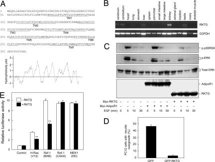
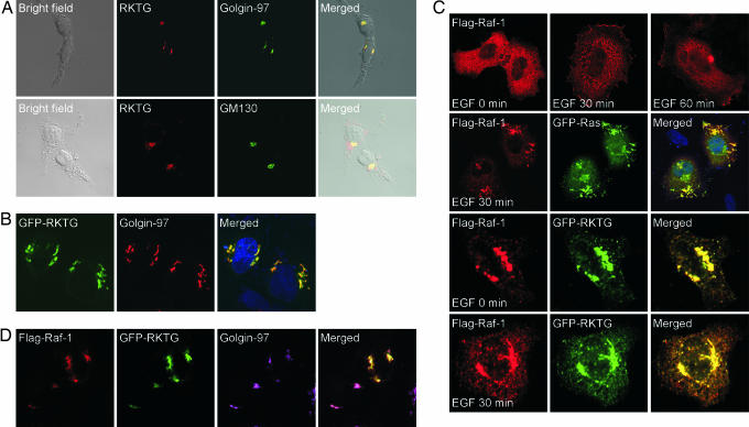
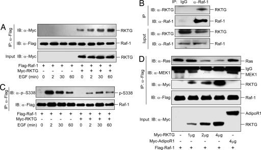
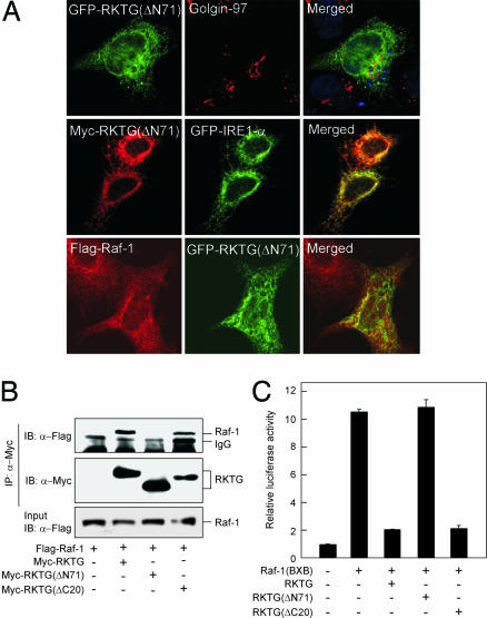
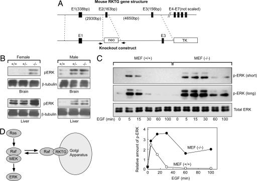
Subcellular compartmentalization has become an important theme in cell signaling such as spatial regulation of Ras by RasGRP1 and MEK/ERK by Sef. Here, we report spatial regulation of Raf kinase by RKTG (Raf kinase trapping to Golgi). RKTG is a seven-transmembrane protein localized at the Golgi apparatus. RKTG expression inhibits EGF-stimulated ERK and RSK phosphorylation, blocks NGF-mediated PC12 cell differentiation, and antagonizes Ras- and Raf-1-stimulated Elk-1 transactivation. Through interaction with Raf-1, RKTG changes the localization of Raf-1 from cytoplasm to the Golgi apparatus, blocks EGF-stimulated Raf-1 membrane translocation, and reduces the interaction of Raf-1 with Ras and MEK1. In RKTG-null mice, the basal ERK phosphorylation level is increased in the brain and liver. In RKTG-deleted mouse embryonic fibroblasts, EGF-induced ERK phosphorylation is enhanced. Collectively, our results reveal a paradigm of spatial regulation of Raf kinase by RKTG via sequestrating Raf-1 to the Golgi apparatus and thereby inhibiting the ERK signaling pathway.


Conflict of interest statement

The authors declare no conflict of interest.

Figures

Fig. 1.
Fig. 1.
Structure, tissue expression, and effect on ERK signaling of RKTG. (A) Amino acid sequence of human RKTG and hydrophobicity analysis. The locations of predicted seven-transmembrane (TM) domains are underlined. (B) Tissue-expression pattern of RKTG. Total RNA was isolated from different mouse tissues as indicated and used in RT-PCR with primers specific for RKTG and G3PDH. (C) Inhibition of EGF-induced ERK and p90RSK phosphorylation by RKTG. HEK293T cells were transiently transfected with Myc-tagged AdipoR1 or RKTG as indicated and then treated with EGF (100 ng/ml) for various amounts of time. Total cell lysate was used in the detection of phosphorylated ERK, phosphorylated p90RSK, total ERK, and Myc-tagged proteins by Western blotting. (D) RKTG inhibits NGF-induced neuritogenesis in PC12 cells. PC12 cells were transfected with GFP control or GFP-RKTG fusion plasmid. The cells were cultured in the presence of NGF (50 ng/ml) for 48 h and then examined by fluorescence microscopy. Cells with processes longer than twice the diameter of the cell body were considered to be positive for neurite outgrowth. The graph data are shown as the mean ± SD from three independent experiments. (E) Regulation of Elk-1-mediated transcriptional response by RKTG. HEK293T cells were transiently transfected with the constitutively active constructs H-Ras(V12), Raf-1(BXB), Raf-1(CAAX), and MEK1(DD) as indicated. The Elk-1-mediated transcriptional response was analyzed as described (39). A renilla luciferase vector was used to monitor the transfection efficiency. The whole-cell lysate was used in a dual luciferase assay, and the fold change of luciferase activity is shown as the mean ± SD. **, P < 0.01 (by Student's t test).
Fig. 2.
Fig. 2.
Raf-1 colocalizes with RKTG at the Golgi apparatus. (A) Endogenous RKTG is localized at the Golgi. HEK293T cells were stained with affinity-purified anti-RKTG antibody (red) and the Golgi markers Golgin-97 (green) or GM130 (green) as indicated. The cells were fixed and used in confocal analysis. The bright fields are also shown to reveal the overall structure of the cells. (B) Ectopically expressed RKTG is localized at the Golgi. HeLa cells were transfected with GFP-RKTG fusion plasmid. The Golgi was labeled by Golgin-97 (red), and the cell nuclei were stained with Hoechest 33342 (blue). (C) Colocalization of RKTG with Raf-1. HeLa cells were transfected with the plasmid as indicated and followed by serum starvation. EGF treatment induced membrane localization of Raf-1 (first row), as well as formation of membrane ruffles that contained Ras and Raf-1 (second row). RKTG coexpression changed the localization of Raf-1 from cytosol to the Golgi in the absence (third row) or presence (fourth row) of EGF. The change of the subcellular localization of Raf-1 by RKTG blocked membrane translocation of Raf-1 upon EGF treatment (bottommost row). (D) Confocal immunofluorescence microscopy of HeLa cells with triple colors. The cells were transfected with Flag-tagged Raf-1 and GFP-RKTG fusion construct and then labeled with a polyclonal antibody against Flag-tagged Raf-1 (red) and a monoclonal antibody against Golgin-97 (purple). Note that the merged image shows a significant overlapping of the three signals.
Fig. 3.
Fig. 3.
Interaction of RKTG with Raf-1. (A) Interaction of overexpressed RKTG with Raf-1. HEK293T cells were transiently transfected with Flag-tagged Raf-1 and Myc-tagged RKTG as indicated. After transfection, the cells were serum-starved for 16 h and then treated with EGF (100 ng/ml) for the indicated times. The cell lysate was used in immunoprecipitation (IP) and immunoblotting (IB) with the antibodies as indicated. (B) Interaction of endogenous RKTG with endogenous Raf-1. Total cell lysates of HEK293T cells were subjected to immunoprecipitation with an anti-Raf-1 antibody followed by immunoblotting with an antibody against human RKTG. Mouse IgG was used as a negative control. (C) RKTG inhibits Raf-1 activation. HEK293T cells were transiently transfected with Flag-tagged Raf-1 and Myc-tagged RKTG. The Flag-tagged Raf-1 was immunoprecipitated after EGF stimulation for indicated times. Immunoblotting was performed by using the antibodies as indicated. (D) RKTG interferes with the interaction of Raf-1 with Ras and MEK1. HEK293T cells were transiently transfected with the plasmid as indicated. The Flag-tagged Raf-1 was immunoprecipitated, and the Raf-1-associated RKTG, Ras, and MEK1 were analyzed by immunoblotting.
Fig. 4.
Fig. 4.
RKTG N terminus is needed for interaction with Raf-1. (A) Colocalization studies of RKTGΔN71 in HeLa cells. (Top) GFP-RKTGΔN71 fusion protein with the Golgi marker Golgin-97. (Middle) Myc-tagged RKTGΔN71 with an ER marker, GFP-IRE1-α. (Bottom) GFP-RKTGΔN71 with Flag-tagged Raf-1. (B) Interaction of RKTG truncation mutants with Raf-1. HEK293 cells were transfected with the plasmids as indicated, and the cell lysate was used in immunoprecipitation (IP) and immunoblotting (IB) with the indicated antibodies. (C) Effects of the N- and C-terminal RKTG deletions on Raf-1(BXB)-induced Elk-1 transactivation. The luciferase assay was performed as described in Fig. 1E.
Fig. 5.
Fig. 5.
Studies with RKTG knockout mouse. (A) Schematic illustration of the mouse RKTG gene structure and the RKTG knockout construct. The seven exons (E1–E7) are marked, and the nucleotide lengths for some of the exons and introns are also shown. Homologous recombination (in the regions marked by dotted lines) would cause deletion of exon 2. The arrows indicate the position of PCR primers used for genotyping the mouse. (B) The basal ERK phosphorylation level is increased in tissues from RKTG-deleted mouse. Brain and liver were isolated from both male and female mice. The basal phosphorylation level of ERK and the loaded protein levels were determined by Western blotting as indicated. (C) EGF-induced ERK phosphorylation is enhanced and prolonged in RKTG-deleted MEF. MEFs isolated from the mouse embryos were cultured in serum-starved medium for 6 h and then treated with EGF treatment (100 ng/ml). The total cell lysate was used in immunoblotting. Both short and long exposures for ERK phosphorylation are shown. The relative amount of ERK phosphorylation as compared with total ERK level is shown in the graph. (D) A model for the function of RKTG. RKTG interacts with cytoplasmic Raf-1 and sequesters Raf-1 in the Golgi apparatus, thereby attenuating the signaling from Ras to MEK and ERK.

Similar articles

Cited by

References

    1. Wellbrock C, Karasarides M, Marais R. Nat Rev Mol Cell Biol. 2004;5:875–885. - PubMed
    1. Kolch W. Nat Rev Mol Cell Biol. 2005;6:827–837. - PubMed
    1. Rapp UR, Gotz R, Albert S. Cancer Cell. 2006;9:9–12. - PubMed
    1. Mor A, Philips MR. Annu Rev Immunol. 2006;24:771–800. - PubMed
    1. Harding A, Tian T, Westbury E, Frische E, Hancock JF. Curr Biol. 2005;15:869–873. - PubMed

Publication types

MeSH terms