

Bfl-1/A1 functions, similar to Mcl-1, as a selective tBid and Bak antagonist
- PMID: 17724464
- PMCID: PMC2880719
- DOI: 10.1038/sj.onc.1210771
Bfl-1/A1 functions, similar to Mcl-1, as a selective tBid and Bak antagonist
Abstract
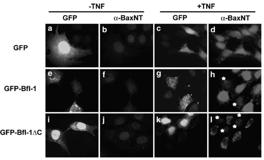
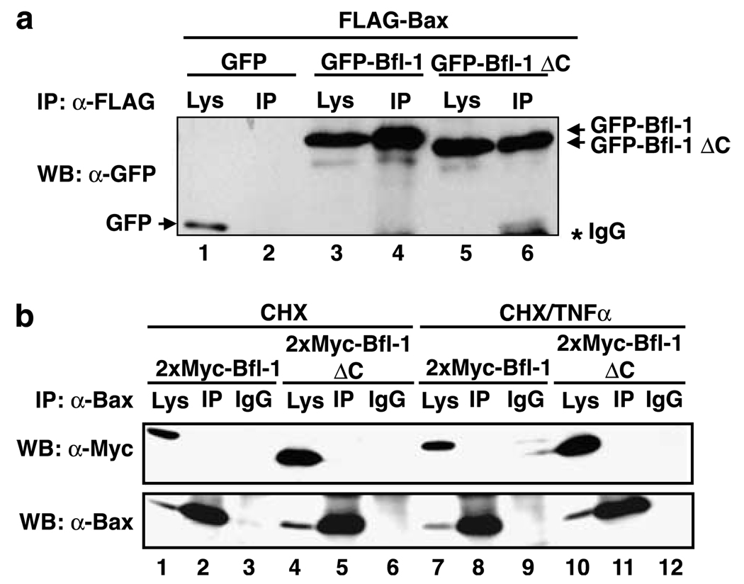
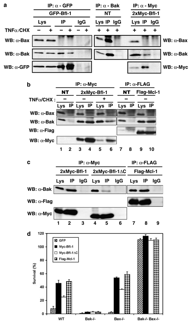
The prosurvival Bcl-2-family member Bfl-1/A1 is a transcriptional target of nuclear factor-kappaB (NF-kappaB) that is overexpressed in many human tumors and is a means by which NF-kappaB inhibits apoptosis, but its mode of action is controversial. To better understand how Bfl-1 functions, we investigated its interaction with proapoptotic multidomain proteins Bax and Bak, and the BH3-only proteins Bid and tBid. We demonstrate that in living cells Bfl-1 selectively interacts with Bak and tBid, but not with Bax or Bid. Bfl-1/Bak interaction is functional as Bfl-1 suppressed staurosporine (STS)-induced apoptosis in wild-type and Bax-deficient cells, but not in Bak-/- cells. We also show that Bfl-1 blocks tumor necrosis factor-alpha (TNFalpha)-induced activation of Bax indirectly, via association with tBid. C-terminal deletion decreased Bfl-1's interaction with Bak and tBid and reduced its ability to suppress Bak- and tBid-mediated cell death. These data indicate that Bfl-1 utilizes different mechanisms to suppress apoptosis depending on the stimulus. Bfl-1 associates with tBid to prevent activation of proapoptotic Bax and Bak, and it also interacts directly with Bak to antagonize Bak-mediated cell death, similar to Mcl-1. Thus, part of the protective function of NF-kappaB is to induce Mcl-1-like activity by upregulating Bfl-1.
Figures


Similar articles
-
Bcl-2 family member Bfl-1/A1 sequesters truncated bid to inhibit is collaboration with pro-apoptotic Bak or Bax.J Biol Chem. 2002 Jun 21;277(25):22781-8. doi: 10.1074/jbc.M201469200. Epub 2002 Apr 19. J Biol Chem. 2002. PMID: 11929871
-
Mcl-1 interacts with truncated Bid and inhibits its induction of cytochrome c release and its role in receptor-mediated apoptosis.J Biol Chem. 2006 Mar 3;281(9):5750-9. doi: 10.1074/jbc.M505688200. Epub 2005 Dec 27. J Biol Chem. 2006. PMID: 16380381
-
Proapoptotic Bak is sequestered by Mcl-1 and Bcl-xL, but not Bcl-2, until displaced by BH3-only proteins.Genes Dev. 2005 Jun 1;19(11):1294-305. doi: 10.1101/gad.1304105. Epub 2005 May 18. Genes Dev. 2005. PMID: 15901672 Free PMC article.
-
Pro-apoptotic cascade activates BID, which oligomerizes BAK or BAX into pores that result in the release of cytochrome c.Cell Death Differ. 2000 Dec;7(12):1166-73. doi: 10.1038/sj.cdd.4400783. Cell Death Differ. 2000. PMID: 11175253 Review.
-
A1/Bfl-1 in leukocyte development and cell death.Exp Cell Res. 2012 Jul 1;318(11):1291-303. doi: 10.1016/j.yexcr.2012.01.021. Epub 2012 Feb 4. Exp Cell Res. 2012. PMID: 22342458 Free PMC article. Review.
Cited by
-
Mechanisms of action of Bcl-2 family proteins.Cold Spring Harb Perspect Biol. 2013 Apr 1;5(4):a008714. doi: 10.1101/cshperspect.a008714. Cold Spring Harb Perspect Biol. 2013. PMID: 23545417 Free PMC article. Review.
-
µ-Calpain conversion of antiapoptotic Bfl-1 (BCL2A1) into a prodeath factor reveals two distinct alpha-helices inducing mitochondria-mediated apoptosis.PLoS One. 2012;7(6):e38620. doi: 10.1371/journal.pone.0038620. Epub 2012 Jun 20. PLoS One. 2012. PMID: 22745672 Free PMC article.
-
GM-CSF Protects Macrophages from DNA Damage by Inducing Differentiation.Cells. 2022 Mar 9;11(6):935. doi: 10.3390/cells11060935. Cells. 2022. PMID: 35326386 Free PMC article.
-
The human pancreatic islet transcriptome: expression of candidate genes for type 1 diabetes and the impact of pro-inflammatory cytokines.PLoS Genet. 2012;8(3):e1002552. doi: 10.1371/journal.pgen.1002552. Epub 2012 Mar 8. PLoS Genet. 2012. PMID: 22412385 Free PMC article.
-
High-quality NMR structure of human anti-apoptotic protein domain Mcl-1(171-327) for cancer drug design.PLoS One. 2014 May 2;9(5):e96521. doi: 10.1371/journal.pone.0096521. eCollection 2014. PLoS One. 2014. PMID: 24789074 Free PMC article.
References
-
- Breitschopf K, Zeiher AM, Dimmeler S. Ubiquitin-mediated degradation of the proapoptotic active form of bid. A functional consequence on apoptosis induction. J Biol Chem. 2000;275:21648–21652. - PubMed
-
- Certo M, Del Gaizo Moore V, Nishino M, Wei G, Korsmeyer S, Armstrong SA, et al. Mitochondria primed by death signals determine cellular addiction to anti-apoptotic BCL-2 family members. Cancer Cell. 2006;9:351–365. - PubMed
-
- Chen L, Willis SN, Wei A, Smith BJ, Fletcher JI, Hinds MG, et al. Differential targeting of prosurvival Bcl-2 proteins by their BH3-only ligands allows complementary apoptotic function. Mol Cell. 2005;17:393–403. - PubMed
-
- Cheng EH, Wei M, Weiler S, Flavell RA, Mak TW, Lindsten T, et al. BCL-2, BCL-XL sequester BH3 domain-only molecules preventing BAX- and BAK-mediated mitochondrial apoptosis. Mol Cell. 2001;8:705–711. - PubMed
-
- Clohessy JG, Zhuang J, de Boer J, Gil-Gomez G, Brady HJ. Mcl-1 interacts with truncated Bid and inhibits its induction of cytochrome c release and its role in receptor-mediated apoptosis. J Biol Chem. 2006;281:5750–5759. - PubMed
Publication types
MeSH terms
Substances
Grants and funding
LinkOut - more resources
Full Text Sources
Research Materials
