Comparative Study
. 2007 Dec 1;408(2):231-40.
doi: 10.1042/BJ20071052.

Identification of a region on hypoxia-inducible-factor prolyl 4-hydroxylases that determines their specificity for the oxygen degradation domains

Affiliations
Comparative Study

Identification of a region on hypoxia-inducible-factor prolyl 4-hydroxylases that determines their specificity for the oxygen degradation domains

Diego Villar et al. Biochem J. .

Abstract

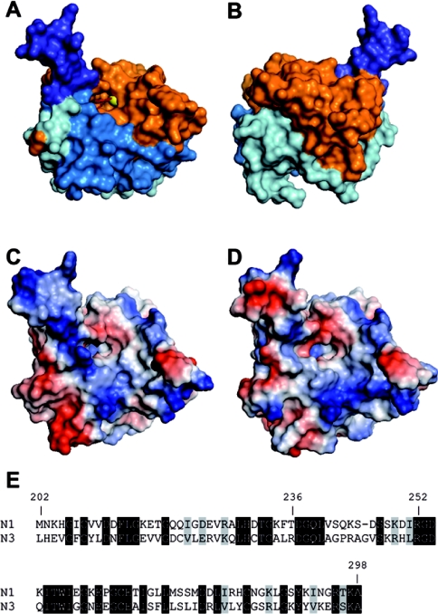
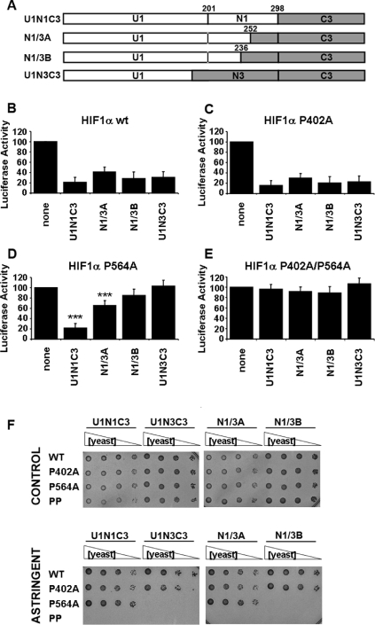
HIFs [hypoxia-inducible (transcription) factors] are essential for the induction of an adaptive gene expression programme under low oxygen partial pressure. The activity of these transcription factors is mainly determined by the stability of the HIFalpha subunit, which is regulated, in an oxygen-dependent manner, by a family of three prolyl 4-hydroxylases [EGLN1-EGLN3 (EGL nine homologues 1-3)]. HIFalpha contains two, N- and C-terminal, independent ODDs (oxygen-dependent degradation domains), namely NODD and CODD, that, upon hydroxylation by the EGLNs, target HIFalpha for proteasomal degradation. In vitro studies indicate that each EGLN shows a differential preference for ODDs, However, the sequence determinants for such specificity are unknown. In the present study we showed that whereas EGLN1 and EGLN2 acted upon any of these ODDs to regulate HIF1alpha protein levels and activity in vivo, EGLN3 only acted on the CODD. With the aim of identifying the region within EGLNs responsible for their differential substrate preference, we investigated the activity and binding pattern of different EGLN deletions and chimaeric constructs generated by domain swapping between EGLN1 and EGLN3. These studies revealed a region of 97 residues that was sufficient to confer the characteristic substrate binding observed for each EGLN. Within this region, we identified the minimal sequence (EGLN1 residues 236-252) involved in substrate discrimination. Importantly, mapping of these sequences on the EGLN1 tertiary structure indicates that substrate specificity is determined by a region relatively remote from the catalytic site.


Figures

Figure 1
Figure 1. Effect of EGLN expression on HIF1α protein levels and activity
(A) HeLa cells were transfected with an HRE-driven reporter construct together with a plasmid encoding for HIF1α (none) or a combination of plasmids encoding for HIF1α and the indicated EGLN enzymes. The graphs show the normalized average reporter activity obtained in three independent experiments (n=3). The HIF1α and α-tubulin (loading control) protein levels were determined in HeLa cell cultures transfected with the indicated combination of plasmids detailed above. (BD) Cells were treated as in (A), except that P402A (B), P564A (C) or P402A/P564A (D) mutant forms of HIF1α were used for the assays. The asterisks (***) indicate that the mean values for EGLN1 and EGLN2 are significantly different (P<0.001) from those obtained for EGLN3. (E) Yeast cells were transformed with AD–HIF1α constructs together with DBD–EGLN1, DBD–EGLN2 or DBD–EGLN3 proteins. Serial dilutions of transformed clones were grown on plates lacking leucine and tryptophan (CONTROL, no interaction required between fusion proteins for yeast growth) or lacking leucine, tryptophan, histidine and adenine (INTERACTION, the interaction between fusion proteins is required for yeast growth). WT, full-length wild-type HIF1α; P402A, HIF1α P402A mutant (intact CODD); P564A, HIF1α P564A mutant (intact NODD); PP, HIF1α P402A/P564A mutant. The results shown are representative for at least three independent experiments. Bkgr, luciferase activity observed in cells transfected with the reporter construct alone; Tub., α-tubulin; WB, Western blot.
Figure 2
Figure 2. Sequence conservation among EGLNs paralogues
Human EGLN1 (NP_071344) was pairwise-aligned with EGLN3 (NP_071356) or EGLN2 (NP_444274) protein sequences using the BLAST algorithm [29]. The graph represents the percentage of sequence identity within a 20 residue sliding window. Positions correspond to EGLN1 sequence. Diagrams above the graph represent the aligned EGLN sequences. Boxes correspond to regions of none (U1/U2), moderate (N1–N3) or high (C1–C3) sequence conservation that were swapped for the generation of chimaeras. Black arrowheads indicate the location of the conserved triad His-Asp-His of residues involved in iron binding at the catalytic centre. Numbers indicate amino acid residue positions.
Figure 3
Figure 3. Effect of different N-terminal deletions on EGLN1 activity
(A) Schematic diagram representing the constructs generated by deletion of the indicated regions of EGLN1. (B) HeLa cells were transfected with an HRE-driven reporter construct, together with a plasmid encoding for HIF1α (none) or a combination of plasmids encoding for HIF1α and the indicated EGLN1 forms. ΔU1, truncated EGLN1 lacking residues 1–177. ΔEGLN1, truncated EGLN1 lacking residues 76–177. The results are normalized average reporter activities obtained in three independent experiments (n=3). The HIF1α and α-tubulin (loading control) protein levels were determined in HeLa cell cultures transfected with the indicated combination of plasmid as detailed above. (CE) Cells were treated as in (A), except that P402A (C), P564A (D) or P402A/P564A (E) mutant forms of HIF1α were used for the assays. (F) Yeast cells were transformed with AD–HIF1α constructs together with wild-type DBD–EGLN1 or the indicated deletion mutants fused to the DBD. Serial dilutions of transformed clones were grown on plates lacking leucine and tryptophan (CONTROL) or plates lacking leucine, tryptophan and histidine (INTERACTION). The results shown are representative for at least three independent experiments. Abbreviations and symbols are as described for Figure 1.
Figure 4
Figure 4. Effect of chimaeric EGLN constructs on HIF1α protein levels and activity
(A) Schematic diagram representing the chimaeric constructs generated by exchange of the indicated regions between EGLN1 and EGLN3. (B) HeLa cells were transfected with an HRE-driven reporter construct together with a plasmid encoding for HIF1α (none) or a combination of plasmids encoding for HIF1α and the indicated wild-type or chimaeric enzymes. A description of the domain structure of the chimerical constructs structure is given in Figure 2. The graphs represent the normalized average reporter activity obtained in three independent experiments (n=3). The asterisks (***) indicate that mean values for those constructs are significantly different (P<0.001) from those obtained for EGLN3. The HIF1α and α-tubulin (loading control) protein levels were determined in HeLa cell cultures transfected with the indicated combination of plasmids as detailed above. (CE) Cells were treated as in (A), except that P402A (C), P564A (D) or P402A/P564A (E) mutant forms of HIF1α were used for the assays. (F) Yeast cells were transformed with AD–HIF1α constructs together with DBD–EGLN1 or DBD–EGLN3 proteins. Serial dilutions of transformed clones were grown on plates lacking leucine and tryptophan (CONTROL) or plates lacking leucine, tryptophan, histidine and adenine (INTERACTION). The results shown are representative for at least three independent experiments. The abbreviations and symbols are as described for Figure 1.
Figure 5
Figure 5. Localization and electrostatic potential of the N-region on EGLN1 structure and EGLN3 model
(A,B and D) Images of the molecular surface of EGLN1 (Protein Data Bank 2G19). The colour code in (A) and (B) was as follows: cyan, residues 188–236; dark blue, residues 237–252; light blue, residues 253–298; and orange, residues 297–403. The wire representation of the 2-oxoglutarate competitive inhibitor compound A is show in red, and the iron atom is represented as a yellow sphere. (A) Front view of the molecule showing the deep catalytic site; (B) 180° rotation of the molecule depicted in (A); (C and D) representation of the qualitative electrostatic surface potential (PyMol) for the EGLN1 structure (D) and a EGLN3 model (C). Shades of red and blue are used to represent negative and positive potentials respectively; (E) alignment of human EGLN1 and EGLN3 sequences. The numbers correspond to residue positions according to the EGLN1 sequence. Identical residues are shown on a black background and conservative substitutions have a grey background.
Figure 6
Figure 6. Effect of N-terminal region chimaeric constructs on HIF1α activity
(A) Schematic diagram representing the chimaeric constructs generated by exchange of the indicated regions between EGLN1 and EGLN3. Numbers indicate amino acid residue positions relative to the EGLN1 sequence. (B) HeLa cells were transfected with an HRE-driven reporter construct together with a plasmid encoding for HIF1α (none) or a combination of plasmids encoding for HIF1α and the indicated chimaeric enzymes. Results are normalized average reporter activities obtained in four independent experiments (n=4). (CE) Cells were treated as in (A), except that P402A (C), P564A (D) or P402A/P564A (E) mutant forms of HIF1α were used for the assays. The asterisks (***) indicate that mean values for N1/3A and U1N1C3 are significantly different (P<0.001) from those obtained for U1N3C3. Differences between N1/3B and U1N3C3 were not statistically significant (P>0.05). (F) Yeast cells were transformed with AD–HIF1α constructs together with DBD–EGLN chimaeric proteins. Serial dilutions of transformed clones were grown on plates lacking leucine and tryptophan (CONTROL) or plates lacking leucine, tryptophan, histidine and adenine (INTERACTION). The results shown are representative for two independent experiments. The abbreviations and symbols used are as described in for Figure 1.

Comment in

Similar articles

Cited by

References

    1. Wenger R. H., Stiehl D. P., Camenisch G. Integration of oxygen signaling at the consensus HRE. Sci. STKE. 2005;306:re12. - PubMed
    1. Schofield C. J., Ratcliffe P. J. Oxygen sensing by HIF hydroxylases. Nat. Rev. Mol. Cell Biol. 2004;5:343–354. - PubMed
    1. Ivan M., Kondo K., Yang H., Kim W., Valiando J., Ohh M., Salic A., Asara J. M., Lane W. S., Kaelin W. G., Jr HIFα targeted for VHL-mediated destruction by proline hydroxylation: implications for O2 sensing. Science. 2001;292:464–468. - PubMed
    1. Jaakkola P., Mole D. R., Tian Y. M., Wilson M. I., Gielbert J., Gaskell S. J., Kriegsheim A., Hebestreit H. F., Mukherji M., Schofield C. J., et al. Targeting of HIF-α to the von Hippel–Lindau ubiquitylation complex by O2-regulated prolyl hydroxylation. Science. 2001;292:468–472. - PubMed
    1. Epstein A. C., Gleadle J. M., McNeill L. A., Hewitson K. S., O'Rourke J., Mole D. R., Mukherji M., Metzen E., Wilson M. I., Dhanda A., et al. C. elegans EGL-9 and mammalian homologs define a family of dioxygenases that regulate HIF by prolyl hydroxylation. Cell. 2001;107:43–54. - PubMed

Publication types

MeSH terms

Substances