

Overexpression of the autoantigen IA-2 puts beta cells into a pre-apoptotic state: autoantigen-induced, but non-autoimmune-mediated, tissue destruction
- PMID: 17725654
- PMCID: PMC2219291
- DOI: 10.1111/j.1365-2249.2007.03455.x
Overexpression of the autoantigen IA-2 puts beta cells into a pre-apoptotic state: autoantigen-induced, but non-autoimmune-mediated, tissue destruction
Abstract
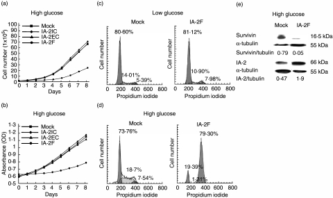
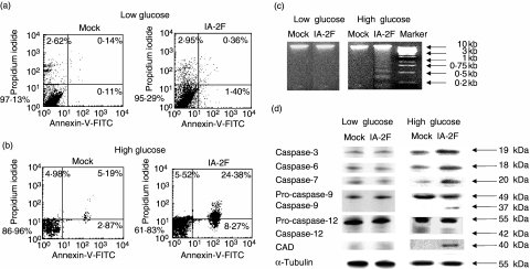
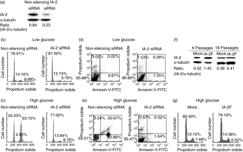
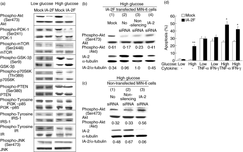
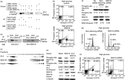
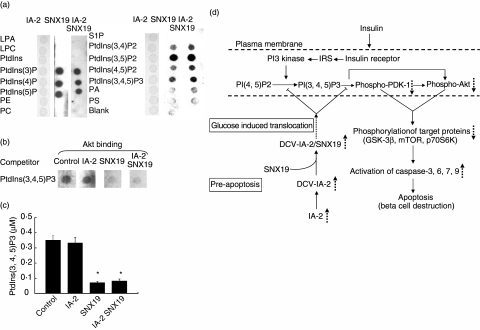
IA-2 is a major autoantigen in type 1 diabetes and autoantibodies to it have become important diagnostic and predictive markers. IA-2 also is an intrinsic transmembrane component of dense core secretory vesicles and knock-out studies showed that IA-2 is a regulator of insulin secretion. Here we show that overexpression of IA-2 puts mouse insulinoma MIN-6 beta cells into a pre-apoptotic state and that exposure to high glucose results in G2/M arrest and apoptosis. Molecular study revealed a decrease in phosphoinositide-dependent kinase (PDK)-1 and Akt/protein kinase B (PKB) phosphorylation. Treatment of IA-2-transfected cells with IA-2 siRNA prevented both G2/M arrest and apoptosis and increased Akt/PKB phosphorylation. A search for IA-2 interacting proteins revealed that IA-2 interacts with sorting nexin (SNX)19 and that SNX19, but not IA-2, inhibits the conversion of PtdIns(4,5)P2 to PtdIns(3,4,5)P3 and thereby suppresses the phosphorylation of proteins in the Akt signalling pathway resulting in apoptosis. We conclude that IA-2 acts through SNX19 to initiate the pre-apoptotic state. Our findings point to the possibility that in autoimmune diseases, tissue destruction may be autoantigen-induced, but not necessarily immunologically mediated.
Figures
 , increase,
  , decrease,
  , inhibition,
  , translocation.
Similar articles
-
Rosiglitazone and PPAR-gamma overexpression protect mitochondrial membrane potential and prevent apoptosis by upregulating anti-apoptotic Bcl-2 family proteins.J Cell Physiol. 2009 Jul;220(1):58-71. doi: 10.1002/jcp.21730. J Cell Physiol. 2009. PMID: 19229877
-
Rictor and integrin-linked kinase interact and regulate Akt phosphorylation and cancer cell survival.Cancer Res. 2008 Mar 15;68(6):1618-24. doi: 10.1158/0008-5472.CAN-07-5869. Cancer Res. 2008. PMID: 18339839
-
[shRNA-mediated insulin-like growth factor I receptor gene silencing inhibits cell proliferation, induces cell apoptosis, and suppresses tumor growth in non-small cell lung cancer: in vitro and in vivo experiments].Zhonghua Yi Xue Za Zhi. 2007 Jun 5;87(21):1506-9. Zhonghua Yi Xue Za Zhi. 2007. PMID: 17785094 Chinese.
-
Altered structure of autoantigens during apoptosis.Rheum Dis Clin North Am. 2004 Aug;30(3):455-71, vii. doi: 10.1016/j.rdc.2004.04.012. Rheum Dis Clin North Am. 2004. PMID: 15261336 Review.
-
B cell biology, apoptosis, and autoantibodies to phospholipids.Thromb Res. 2004;114(5-6):307-19. doi: 10.1016/j.thromres.2004.06.037. Thromb Res. 2004. PMID: 15507260 Review.
Cited by
-
SNX19 restricts endolysosome motility through contacts with the endoplasmic reticulum.Nat Commun. 2021 Jul 27;12(1):4552. doi: 10.1038/s41467-021-24709-1. Nat Commun. 2021. PMID: 34315878 Free PMC article.
-
SNX19 Interacts with Caveolin-1 and Flotillin-1 to Regulate D1R Endocytosis and Signaling.Biomedicines. 2025 Feb 15;13(2):481. doi: 10.3390/biomedicines13020481. Biomedicines. 2025. PMID: 40002894 Free PMC article.
-
Disturbances in the secretion of neurotransmitters in IA-2/IA-2beta null mice: changes in behavior, learning and lifespan.Neuroscience. 2009 Mar 17;159(2):427-37. doi: 10.1016/j.neuroscience.2009.01.022. Neuroscience. 2009. PMID: 19361477 Free PMC article.
-
Proteome profiling of different rat brain regions reveals the modulatory effect of prolonged maternal separation on proteins involved in cell death-related processes.Biol Res. 2021 Feb 8;54(1):4. doi: 10.1186/s40659-021-00327-5. Biol Res. 2021. PMID: 33557947 Free PMC article.
-
Incidental CD8 T cell reactivity against caspase-cleaved apoptotic self-antigens from ubiquitously expressed proteins in islets from prediabetic human leucocyte antigen-A2 transgenic non-obese diabetic mice.Clin Exp Immunol. 2011 Aug;165(2):155-62. doi: 10.1111/j.1365-2249.2011.04420.x. Epub 2011 May 23. Clin Exp Immunol. 2011. PMID: 21605113 Free PMC article.
References
-
- Notkins AL. Immunologic and genetic factors in type 1 diabetes. J Biol Chem. 2002;277:43545–8. - PubMed
-
- Lan MS, Lu J, Goto Y, et al. Molecular cloning and identification of a receptor-type protein tyrosine phosphatase, IA-2, from human insulinoma. DNA Cell Biol. 1994;13:505–14. - PubMed
-
- Lu J, Notkins AL, Lan MS. Isolation, sequence and expression of a novel mouse brain cDNA, mIA-2, and its relatedness to members of the protein tyrosine phosphatase family. Biochem Biophys Res Commun. 1994;204:930–6. - PubMed
-
- Magistrelli G, Toma S, Isacchi A. Substitution of two variant residues in the protein tyrosine phosphatase-like PTP35/IA-2 sequence reconstitutes catalytic activity. Biochem Biophys Res Commun. 1996;227:581–8. - PubMed
MeSH terms
Substances
LinkOut - more resources
Full Text Sources
Miscellaneous
