. 2007 Aug 28:8:37.
doi: 10.1186/1471-2121-8-37.

Identifying genes preferentially expressed in undifferentiated embryonic stem cells

Affiliations

Identifying genes preferentially expressed in undifferentiated embryonic stem cells

Xiajun Li et al. BMC Cell Biol. .

Abstract

Background: The mechanism involved in the maintenance and differentiation of embryonic stem (ES) cells is incompletely understood.

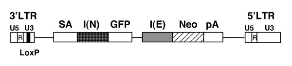
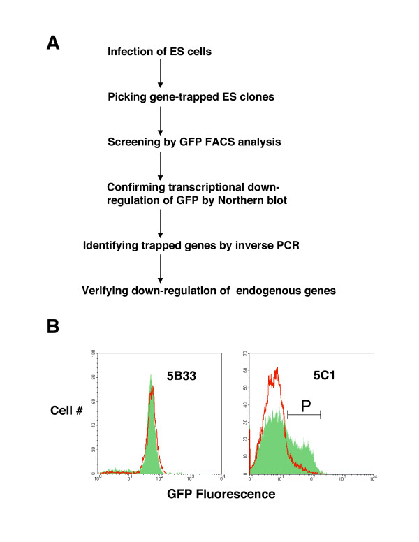
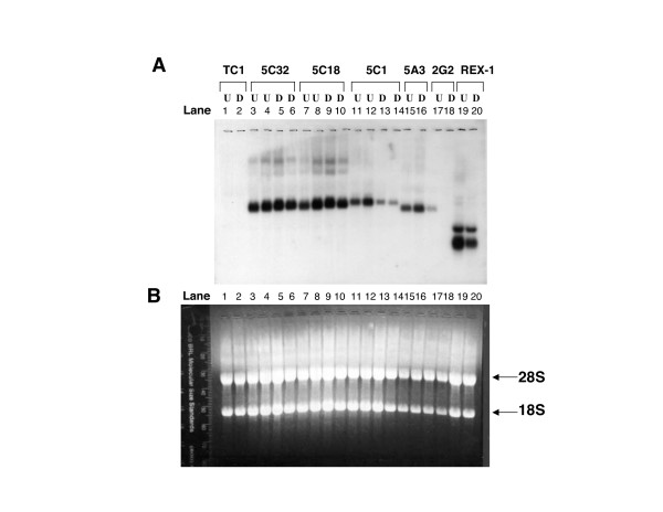
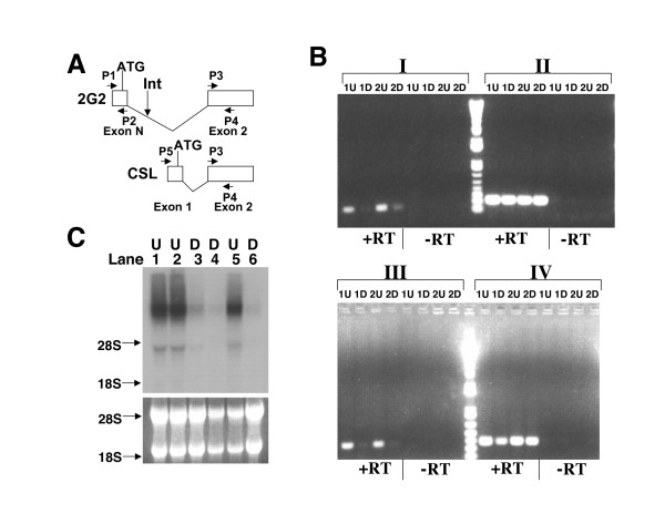
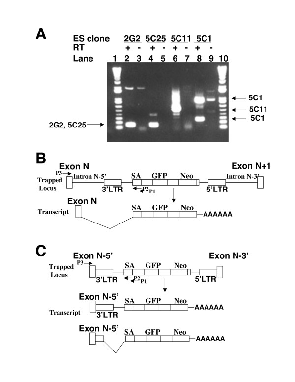
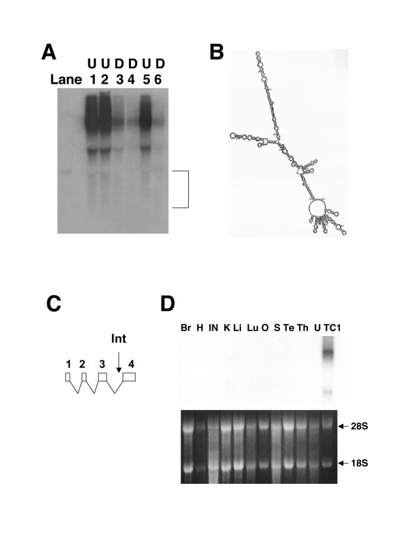
Results: To address this issue, we have developed a retroviral gene trap vector that can target genes expressed in undifferentiated ES cells. This gene trap vector harbors both GFP and Neo reporter genes. G-418 drug resistance was used to select ES clones in which the vector was integrated into transcriptionally active loci. This was then followed by GFP FACS profiling to identify ES clones with reduced GFP fluorescence and, hence, reduced transcriptional activity when ES cells differentiate. Reduced expression of the GFP reporter in six of three hundred ES clones in our pilot screening was confirmed to be down-regulated by Northern blot analysis during ES cell differentiation. These six ES clones represent four different genes. Among the six integration sites, one was at Zfp-57 whose gene product is known to be enriched in undifferentiated ES cells. Three were located in an intron of a novel isoform of CSL/RBP-J kappa which encodes the key transcription factor of the LIN-12/Notch pathway. Another was inside a gene that may encode noncoding RNA transcripts. The last integration event occurred at a locus that may harbor a novel gene.

Conclusion: Taken together, we demonstrate the use of a novel retroviral gene trap vector in identifying genes preferentially expressed in undifferentiated ES cells.


Figures

Figure 1
Figure 1
Diagram of the gene trap vector eGeoN/E+pA. Neo, Neo resistance gene; GFP, cDNA encoding green fluorescent protein; pA, polyA addition signal derived from the gene of bovine growth hormone (bGH); SA, splice acceptor sequence present in intron 2/exon 3 of Bcl-2; I(E), IRES sequence from encephalomyocarditis virus (EMCV); I(N), IRES sequence from the 5' UTR region of the NF-kappaB repressing factor (NRF) gene; 3'LTR and 5'LTR, long terminal repeat regions of the RET vector modified from murine Moloney leukemia virus [23]. Both 3'LTR and 5'LTR are comprised of U5, R and U3. The regulatory sequence in the U3 region of 3'LTR was deleted and replaced with a loxP site which could be recognized by Cre recombinase.
Figure 2
Figure 2
G-418 drug resistance and GFP fluorescence were used to isolate the trapped ES clones whose trapped genes are down-regulated during ES cell differentiation. (A) Flow chart for the experimental procedure.(B) GFP FACS profiling of the ES clones. FACS plots of the ES clone 5B33 (left panel) and ES clone 5C1 (right panel) are shown with both the undifferentiated populations of cells (the green filled histograms) and differentiated populations of cells (the red unfilled histograms). Undifferentiated ES cells were grown in the presence of LIF and feeder fibroblast cells and the differentiated cells were grown without LIF and feeder fibroblast cells. "P" marks the GFP-positive population of ES cells present in the undifferentiated cells of the ES clone 5C1.
Figure 3
Figure 3
Northern blot analysis of the candidate ES clones. (A) The exposed X-ray film of the Northern blot with a GFP cDNA fragment as the probe. U, Undifferentiated ES cell population. D, Differentiated ES cell population. TC1, wild-type ES cells. REX-1, an ES clone with the REX1::EGFP reporter transgene. 5C32, 5C18, 5C1, 5A3 and 2G2 are gene-trapped ES clones isolated in this study. (B) The RNA-agarose gel under an ultraviolet (UV) lamp. Approximately 10 μg of total RNA was loaded for the U and D samples of the control samples and each ES clone, except that 5 μg of total RNA was applied for the ES clone 5A3 and 2G2. The positions of 18S and 28S ribosomal RNAs on the agrose gel are marked.
Figure 5
Figure 5
Verifying the down-regulation of the endogenous genes during ES cell differentiation. (A) Diagrams are shown for the 5' portion of the novel embryonic isoform (upper, 2G2) as well as the most common isoform (lower, CSL) of CSL/RBP-Jkappa. The first exon (Exon N) of the novel embryonic isoform is located upstream of the second exon (Exon 2) which is common to all known isoforms. The predicted initiation codon "ATG" is indicated above the first exons and the primers (P1, P2, P3, P4 and P5) used in the semi-qauntitative PCR analysis below (Figure 5B) are also shown. "Int" stands for the retroviral integration site in the ES clone 2G2, which is located within the first intron of the novel embryonic isoform. Retroviral integration occurred in the same intron for two other ES clone 5C25 and 6B13. (B) Verification of the reduced expression of the novel early embryonic isoform of CSL/RBP-Jkappa in the differentiated cells (D) versus undifferentiated (U) cells by quantitative RT-PCR analysis. 1U and 2U, two independent total RNA samples from the undifferentiated wild-type ES cells. 1D and 2D, two independent total RNA samples from the differentiated cells. Equal amounts of total RNA was applied to every PCR reaction in each primer set. Four different primer sets (I, II, III, IV) were used in this quantitative PCR analysis. I, both primers (P1 and P2) are derived from the first exon unique to this novel isoform (Figure 5A). II, both primers (P3 and P4) are common to all the isoforms of CSL/RBP-Jkappa. III, the forward primer (P1) is derived from the first exon and the reverse primer (P4) is complementary in sequence to the second exon, which is common to all isoforms of CSL/RBP-Jkappa. IV, the forward primer (P5) corresponds to the first exon of the most common isoform of CSL/RBP-Jkappa and the reverse primer (P4) is complementary in sequence to the common second exon. "+RT" indicates that reverse transcriptase (RT) was included in the first-strand cDNA synthesis. "-RT" indicates negative controls in which no reverse transcriptase was added in the first-strand cDNA synthesis. (C) Confirming the down-regulation of the novel gene trapped in the ES clone 5C11 by Northern blot with a gene-specific probe. Total RNA samples were derived from three independent populations (Lanes 1, 2, 5) of the undifferentiated (U) wild-type ES cells and three independent populations (Lanes 3, 4, 6) of the differentiated (D) wild-type ES cells. The gel positions of 18S and 28S ribosomal RNA transcripts are marked with arrows.
Figure 4
Figure 4
The identities of the trapped genes were verified by RT-PCR analysis of the fusion transcripts. (A) RT-PCR products of the four candidate ES clones (2G2, 5C25, 5C11 and 5C1). The primer Xho-Junc (P1), which has a sequence complementary to the junction of the SA site and I(N) sequence in the gene trap vector eGeoN/E+pA, was used for reverse transcription in the first-strand cDNA synthesis. Another primer Bcl-2R2 (P2) complementary to exon 3 of Bcl-2 in eGeoN/E+pA was paired with the gene-specific primer (P3) to amplify the cDNAs derived from the fusion transcripts between the endogenous trapped genes and the gene trap vector. The positions of these primers (P1, P2, P3) with respect to the gene trap vector and the trapped endogenous genes are indicated in Figure 4B and Figure 4C. Arrows indicate the agarose gel positions of the expected RT-PCR products. "+", reverse transcriptase (RT) was included in the first-strand cDNA synthesis (Lanes 2, 4, 6, 8). "-", no reverse transcriptase was added to the first-strand cDNA synthesis (Lanes 3, 5, 7, 9). Lane 1 and Lane 11, 1kb plus DNA ladder (Invitrogen/Gibco). (B) Schematic diagrams illustrating the fusion transcripts expressed in the trapped ES clones when the gene trap vector is inserted into an intron of the active endogenous genes (e.g. ES clone 2G2 and 5C25). As shown in the diagrams, the vector is inserted into the Intron N between Exon N and Exon N+1. After the splicing of the fusion transcripts, the splice donor present at the junction of Exon N/Intron N was utilized to join the Exon N directly to the splice acceptor (SA) present in front of the GFP and Neo reporter genes. (C) Schematic diagrams illustrating the fusion transcripts expressed in the trapped ES clones when the gene trap vector is integrated into an exon of the active endogenous genes (e. g. ES clone 5C1, the first exon of Zfp-57). As shown in the diagrams, the vector is integrated into Exon N, which results in the split of Exon N into two parts (Exon N-5' and Exon N-3'). A read-through fusion transcript will be generated or the cryptic splice donor site present in the 3'LTR of the vector is utilized to join the Exon N-5' part and portion of the 3'LTR directly to the splice acceptor (SA) present in front of the GFP and Neo reporter genes.
Figure 6
Figure 6
The novel gene trapped in 5C11 may encode noncoding RNA transcripts with a very restricted expression pattern. (A) Longer exposure (10-day) of the Northern blot as shown in Figure 5B revealed the presence of short transcripts of 5C11. These heterogeneous short transcripts, marked by a bracket, were also down-regulated during ES cell differentiation by comparing Lanes 1, 2, 5 with Lanes 3, 4, 6. (B) Short transcripts of 5C11 were predicted to form very stable secondary structures. The nucleic acid folding program (mfold) was used for the prediction [47]. One predicted folded RNA structure for a full-length cDNA is shown here as an example. (C) A diagram is shown for the exon-intron organization of one of the isoforms of 5C11 transcripts. "Int" stands for the retroviral integration site in the ES clone 5C11. This isoform is predicted to form highly stable secondary structures (Figure 6B). (D) 5C11 transcripts are not detectable in any adult mouse organs by Northern blot. Total RNA samples were derived from the following adult mouse organs: Br, Brain; H, Heart; IN, Intestine; K, Kidney; Li, Liver; Lu, Lung; O, Ovary; S, Spleen; Te, Testis; Th, Thymus; U, Uterus. Total RNA was also derived from wild-type TC1 ES cells (TC1). The gel positions of the ribosomal RNA transcripts were marked.

Similar articles

Cited by

References

    1. McKay R. Stem cells – hype and hope. Nature. 2000;406:361–4. doi: 10.1038/35019186. - DOI - PubMed
    1. Odelberg SJ, Kollhoff A, Keating MT. Dedifferentiation of mammalian myotubes induced by msx1. Cell. 2000;103:1099–109. doi: 10.1016/S0092-8674(00)00212-9. - DOI - PubMed
    1. Terada N, Hamazaki T, Oka M, Hoki M, Mastalerz DM, Nakano Y, Meyer EM, Morel L, Petersen BE, Scott EW. Bone marrow cells adopt the phenotype of other cells by spontaneous cell fusion. Nature. 2002;416:542–5. doi: 10.1038/nature730. - DOI - PubMed
    1. Ying QL, Nichols J, Evans EP, Smith AG. Changing potency by spontaneous fusion. Nature. 2002;416:545–8. doi: 10.1038/nature729. - DOI - PubMed
    1. Scholer HR, Hatzopoulos AK, Balling R, Suzuki N, Gruss P. A family of octamer-specific proteins present during mouse embryogenesis: evidence for germline-specific expression of an Oct factor. Embo J. 1989;8:2543–50. - PMC - PubMed

Publication types

Substances

LinkOut - more resources