. 2007;35(17):5944-53.
doi: 10.1093/nar/gkm641. Epub 2007 Aug 28.

Tissue-dependent paired expression of miRNAs

Affiliations

Tissue-dependent paired expression of miRNAs

Seungil Ro et al. Nucleic Acids Res. 2007.

Abstract

It is believed that depending on the thermodynamic stability of the 5'-strand and the 3'-strand in the stem-loop structure of a precursor microRNA (pre-miRNA), cells preferentially select the less stable one (called the miRNA or guide strand) and destroy the other one (called the miRNA* or passenger strand). However, our expression profiling analyses revealed that both strands could be co-accumulated as miRNA pairs in some tissues while being subjected to strand selection in other tissues. Our target prediction and validation assays demonstrated that both strands of a miRNA pair could target equal numbers of genes and that both were able to suppress the expression of their target genes. Our finding not only suggests that the numbers of miRNAs and their targets are much greater than what we previously thought, but also implies that novel mechanisms are involved in the tissue-dependent miRNA biogenesis and target selection process.


Figures

Figure 1.
Figure 1.
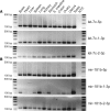
Duplex structures and expression profiles of the 5′-strand and the 3′-strand miRNAs of mir-24 (A–C), mir-194 (D–F) and mir-130a (G–I) in multiple mouse tissues. Both strands of mir-24 (mir-24-5p and -3p) were previously identified in the mouse and human. The 3′-strand of mir-194 (mir-194-3p) and the 5′-strand of mir-130a (mir-130a-5p) were predicted in this study and marked with asterisks (*). RNA sequences for each strand in the core used for designing PCR primers are indicated by arrows. Semi-quantitative PCRs were performed to examine levels of the 5′- and the 3′-strands of each miRNA (B for mir-24-5p and -3p, E for mir-194-5p and -3p and H for mir-130-5p and -3p) in 12 mouse tissues. The cycle numbers were empirically determined to ensure all amplification reactions were in the exponential range. A housekeeping miRNA let-7d-5p was used as a loading control. NTC stands for non-template negative control. A DNA ladder on each side indicates the size of the fragments. Detection of the 5′-strand and the 3′-strand miRNAs of mir-24 (C), mir-194 (F) and mir-130a (I) in six mouse tissues by ribonuclease protection assays are shown. Two controls, probe (probe without RNA sample and without RNase treatment), and no target (sample without RNA sample and with RNase treatment) for each miRNA were included.
Figure 2.
Figure 2.
Expression profiles of miRNA pairs of let-7c and mir-181b transcribed from two loci on different chromosomes. Levels of the 5′- and the 3′-strands of each miRNA in 12 mouse tissues were analyzed using semi-quantitative RT-PCR with cycle numbers empirically determined to be within the exponential range. NTC stands for a non-template control. A DNA ladder on each side indicates the size of the fragments. A housekeeping miRNA let-7c-5p was used as a loading control. (A) Expression profiles of let-7c-5p and two sister 3′-strand miRNAs transcribed from two loci on chromosomes 16 and 15. Note: let-7c-5p transcribed from the two loci displays an identical sequence, whereas the two sister 3′-strand miRNAs transcribed from the two loci are slightly different in their sequences and thus can be distinguished using specific primers in the PCR assays (for sequences see Supplementary Table 8). (B) Expression profiles of mir-181b-5p and the two sister 3′-strand miRNAs transcribed from two loci on chromosomes 1 and 2. Note: mir-181b-5p transcribed from the two loci have an identical sequence, whereas two sister 3′-strand miRNAs derived from the two loci are different in their sequences and thus can be distinguished using specific primers in the PCR assays (for sequences see Supplementary Table 8).
Figure 3.
Figure 3.
Average numbers of target genes predicted for paired miRNAs. Target genes for the 5′- or 3′-strands of paired miRNAs from mouse (86 miRNAs/43 pairs) and human (120 miRNAs/60 pairs) were predicted using the miRanda algorithm with a P-value cutoff at 0.05. Data were retrieved using the miRBase Targets Version 4 (Means ± SD, n = 204).
Figure 4.
Figure 4.
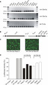
Suppression of target gene expression by a miRNA pair mir-30e-5p and mir-30e-3p. (A) Steady-state expression levels of mir-30e-5p and mir-30e-3p in 12 mouse tissues determined by semi-quantitative RT-PCR analyses. NTC stands for a non-template control. A DNA ladder on each side indicates the size of the fragments. A housekeeping miRNA let-7d-5p was used as a loading control. (B) A schematic map of the mir-30e-target validation vector. The pre-mir-30e (98 bp) with a chimeric intron was inserted in the middle of the eGFP-coding region (eGFPa and eGFPb). Each of the target-binding regions from the six predicted target genes (Akap14, Tshb, Sap30, Mrps30, Npffr2 and Spata19) was inserted into the 3′UTR of the Luciferase gene (Luc2). PSV40, SV40 promoter; Tg, target region; pA, poly (A) signal; PCMV, CMV promoter. (C) GFP-positive HEK-293 cells transfected with mir-30e-5p-Akap14 (left) and mir-30e-3p-Mrps30 (right). Transfected cells were cultured for 24 h before visualization and photography under a fluorescent microscope. (D) Suppression of luciferase activity by interactions between the sister miRNA pair mir-30e-5p and mir-30e-3p and their corresponding targeting sequences at the 3′UTR of Luc2. Luciferase activities (Firefly luciferase and Renilla luciferase) in the cells co-transfected with each of the six target validation plasmids and pRL-CMV were measured. As negative controls, pGL-mir-30eTar (containing no target sequences) and pGL-mir-30e-Gpr137 (containing a let-7d-5p target sequence) were used. The no-protein controls represent samples added with the buffer only and the eGFP controls were those transfected with the eGFP-expressing vector eGFP-pre-miRNA/pDNA3.1 only. The firefly luciferase activity was normalized against the Renilla luciferase. Reduction of the luciferase activity was presented as percentage of levels in the no target samples (Means ± SEM, n = 3).
Figure 5.
Figure 5.
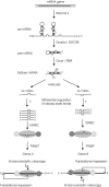
A new ‘Target-Two-Sets-of-Genes-With-One-Pre-miRNA’ model for miRNA biogenesis and target selection. In this model, a miRNA gene is transcribed by RNA polymerase II and is modified in the nucleus into a pri-miRNA, which is further processed to produce an ∼60–70 nt pre-miRNA by the RNase III endonuclease Drosha and DGCR8. The pre-miRNA is then exported to the cytoplasm by Ran-GTP and Exportin-5. A 22-nt miRNA duplex is cleaved from the pre-miRNA by the second RNase III endonuclease complex including Dicer and TRBP. This mature miRNA duplex contains a 5′-strand (5p) miRNA and a sister 3′-strand (3p) miRNA, which is unwound by Helicase. Steady-state levels of each of the miRNA pair are independently regulated by an unknown tissue-specific mechanism. Each strand of the sister miRNA pair can be incorporated into miRISC to become an active miRNA. Since sister miRNAs bound to miRISC possess different sequences, they may target different portions of the same genes or completely different sets of genes to mediate the degradation or translational inhibition of their target mRNAs.

Similar articles

Cited by

References

    1. Ambros V, Bartel B, Bartel DP, Burge CB, Carrington JC, Chen XM, Dreyfuss G, Eddy SR, Griffiths-Jones S, et al. A uniform system for microRNA annotation. RNA. 2003;9:277–279. - PMC - PubMed
    1. Griffiths-Jones S. The microRNA Registry. Nucleic Acids Res. 2004;32:D109–D111. - PMC - PubMed
    1. Griffiths-Jones S, Grocock RJ, van Dongen S, Bateman A, Enright AJ. miRBase: microRNA sequences, targets and gene nomenclature. Nucleic Acids Res. 2006;34:D140–D144. - PMC - PubMed
    1. Kim VN. MicroRNA biogenesis: coordinated cropping and dicing. Nat. Rev. Mol. Cell Biol. 2005;6:376–385. - PubMed
    1. Chen XM. A microRNA as a translational repressor of APETALA2 in Arabidopsis flower development. Science. 2004;303:2022–2025. - PMC - PubMed

Publication types