. 2007;35(17):5913-21.
doi: 10.1093/nar/gkm626. Epub 2007 Aug 28.

Molecular basis of Diamond-Blackfan anemia: structure and function analysis of RPS19

Affiliations

Molecular basis of Diamond-Blackfan anemia: structure and function analysis of RPS19

Lynn A Gregory et al. Nucleic Acids Res. 2007.

Abstract

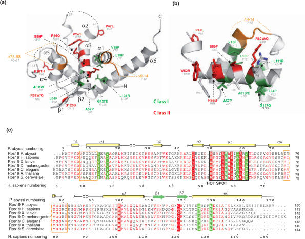
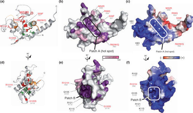
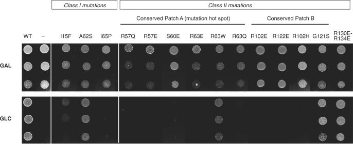
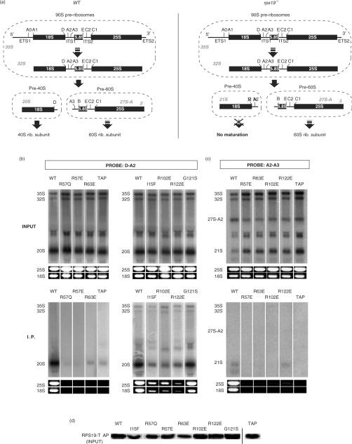
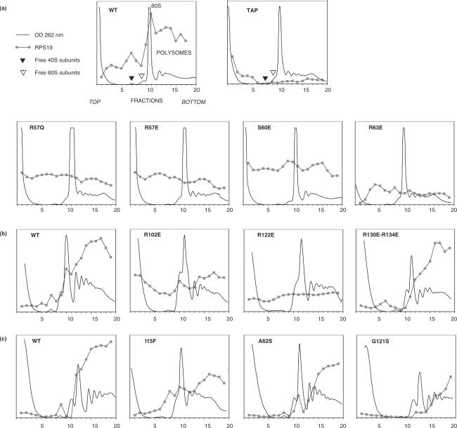
Diamond-Blackfan anemia (DBA) is a rare congenital disease linked to mutations in the ribosomal protein genes rps19, rps24 and rps17. It belongs to the emerging class of ribosomal disorders. To understand the impact of DBA mutations on RPS19 function, we have solved the crystal structure of RPS19 from Pyrococcus abyssi. The protein forms a five alpha-helix bundle organized around a central amphipathic alpha-helix, which corresponds to the DBA mutation hot spot. From the structure, we classify DBA mutations relative to their respective impact on protein folding (class I) or on surface properties (class II). Class II mutations cluster into two conserved basic patches. In vivo analysis in yeast demonstrates an essential role for class II residues in the incorporation into pre-40S ribosomal particles. This data indicate that missense mutations in DBA primarily affect the capacity of the protein to be incorporated into pre-ribosomes, thus blocking maturation of the pre-40S particles.


Figures

Figure 1.
Figure 1.
RPS19 structure and sequence alignment. (a) Overall structure of RPS19 and point mutations found in DBA patients. Residues labeled in green and red correspond to structural residues (class I) and to solvent accessible residues (class II), respectively. Green and red numbers correspond to human numbering. Gray numbers correspond to P. abyssi numbering. Deletions encountered in DBA patients are shown in orange. Dashed lines correspond to disordered loops in the structure. (b) The hot spot of mutations. Mutated residues found in and around the hot spot of mutation are shown as in (a). (c) Sequence alignment of RPS19 orthologs. RPS19 ortholog sequences have been retrieved and aligned using PipeAlign program suite (31). Class I (boxed in green) and class II (boxed in red) residues are displayed. Deletions encountered in DBA patients are show as orange boxes. Secondary sequence elements are shown on top of the sequence.
Figure 2.
Figure 2.
Inter-species conservation and electrostatic surface properties of RPS19. Panels a–c and panels d–f are presented under the same orientation, respectively. (a) and (d) Ribbon diagrams of RPS19. (b) and (e) Surface residue conservation of RPS19. Surface conservation was calculated based on the sequence alignment shown in Figure 1 and using ConSurf server with default parameters (32). Conservation is displayed according to white (non-conserved) to purple (100% conservation). Conserved regions are circled in black. Numbering follows the same rule as in Figure 1. (c) and (f) Electrostatic charge distribution on the surface of RPS19 was calculated using APBS program default parameters (33). Highly conserved and charged areas are boxed.
Figure 3.
Figure 3.
Effect of mutations on RPS19 function in yeast. Capacity of various RPS19 mutants to complement RPS19 expression knockdown was tested in a yeast strain expressing wild-type RPS19 under control of a GAL promoter. Expression of wild-type RPS19 gene is shutdown upon transfer to glucose containing medium whereas transcription of the mutant forms is driven by a constitutive promoter. Cells were spotted at three different densities.
Figure 4.
Figure 4.
Co-immunoprecipitation of precursor and mature ribosomal RNAs with RPS19-TAP. (a) Schematic of pre-rRNA processing in wild-type (top panel) or rps19 deficient cells (lower panel). Letters above pre-rRNAs indicate cleavage sites. (b, c) TAP-tagged wild-type or mutated RPS19A expressed in rps19AΔ/RPS19B yeast cells were isolated with IgG Sepharose beads. Pre-rRNAs in the whole cell extract (‘Input’) and the isolated material (‘I.P.’) were analyzed by northern blot with probes complementary to segment D-A2 (panel B) or to segment A2-A3 (panel C). The mature 18S and 25S rRNAs were detected by ethidium bromide staining. The three panels correspond to independent experiments. In panels A and C, expression of the sole TAP tag was used as a negative control (TAP). The precipitation background level was also evaluated by detecting the 27S-B pre-rRNA with a probe complementary to the ITS2 (data not shown): <0.1% was co-precipitated in panel A and C, <0.3% in panel B, as measured by phosphorimaging. The portion of co-precipitated 20S, 18S and 25S RNAs did not significantly exceed the background value in all panels, except for the wild-type and G121S forms (1–3% of co-precipitation). (d) Levels of TAP-tagged proteins in inputs (corresponding to panel A) were analyzed by western blot with peroxidase anti-peroxidase antibody complexes, which bind to protein A.
Figure 5.
Figure 5.
Analysis of wild-type and mutant RPS19 association to ribosomes on sucrose gradient.Ribosomes from rps19AΔ/RPS19B yeast strains expressing wild-type or mutated forms of RPS19A fused to the TAP tag were separated on sucrose gradient as described in material and methods. RPS19-TAP was detected in gradient fractions on slot blots with peroxidase. Panels a, b and c correspond to three series of experiments. Experiments were performed two or three times with similar results. Expression of the sole TAP tag was used as a negative control (TAP).

Similar articles

Cited by

References

    1. Da Costa L, Willig TN, Fixler J, Mohandas N, Tchernia G. Diamond-Blackfan anemia. Curr. Opin. Pediatr. 2001;13:10–15. - PubMed
    1. Willig TN, Gazda H, Sieff CA. Diamond-Blackfan anemia. Curr. Opin. Hematol. 2000;7:85–94. - PubMed
    1. Gustavsson P, Garelli E, Draptchinskaia N, Ball S, Willig TN, Tentler D, Dianzani I, Punnett HH, Shafer FE, et al. Identification of microdeletions spanning the Diamond-Blackfan anemia locus on 19q13 and evidence for genetic heterogeneity. Am. J. Hum. Genet. 1998;63:1388–1395. - PMC - PubMed
    1. Draptchinskaia N, Gustavsson P, Andersson B, Pettersson M, Willig TN, Dianzani I, Ball S, Tchernia G, Klar J, et al. The gene encoding ribosomal protein S19 is mutated in Diamond-Blackfan anaemia. Nat. Genet. 1999;21:169–175. - PubMed
    1. Leger-Silvestre I, Caffrey JM, Dawaliby R, Alvarez-Arias DA, Gas N, Bertolone SJ, Gleizes PE, Ellis SR. Specific role for yeast homologs of the Diamond Blackfan anemia-associated Rps19 protein in ribosome synthesis. J. Biol. Chem. 2005;280:38177–38185. - PubMed

Publication types