. 2007 Sep 15;120(Pt 18):3200-6.
doi: 10.1242/jcs.014902. Epub 2007 Aug 28.

Cdc42 acts downstream of Bazooka to regulate neuroblast polarity through Par-6 aPKC

Affiliations

Cdc42 acts downstream of Bazooka to regulate neuroblast polarity through Par-6 aPKC

Scott X Atwood et al. J Cell Sci. .

Abstract

Cdc42 recruits Par-6-aPKC to establish cell polarity from worms to mammals. Although Cdc42 is reported to have no function in Drosophila neuroblasts, a model for cell polarity and asymmetric cell division, we show that Cdc42 colocalizes with Par-6-aPKC at the apical cortex in a Bazooka-dependent manner, and is required for Par-6-aPKC localization. Loss of Cdc42 disrupts neuroblast polarity: cdc42 mutant neuroblasts have cytoplasmic Par-6-aPKC, and this phenotype is mimicked by neuroblast-specific expression of a dominant-negative Cdc42 protein or a Par-6 protein that lacks Cdc42-binding ability. Conversely, expression of constitutively active Cdc42 leads to ectopic Par-6-aPKC localization and corresponding cell polarity defects. Bazooka remains apically enriched in cdc42 mutants. Robust Cdc42 localization requires Par-6, indicating the presence of feedback in this pathway. In addition to regulating Par-6-aPKC localization, Cdc42 increases aPKC activity by relieving Par-6 inhibition. We conclude that Cdc42 regulates aPKC localization and activity downstream of Bazooka, thereby directing neuroblast cell polarity and asymmetric cell division.


Figures
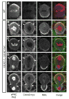
Figure 1
Figure 1. Cdc42 is enriched at the apical neuroblast cortex
(A) Wild-type central brain neuroblasts at 120 h after larval hatching (ALH). Normal apical and basal protein localization is shown with background c-myc staining. (B-E) cdc42-3 central brain neuroblasts at 96 h ALH expressing Cdc42:myc under its native promoter. All stages of mitosis represented. Arrowheads delineate extent of aPKC and Cdc42:myc apical crescents.
Figure 2
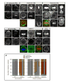
Figure 2. Cdc42 is required for neuroblast polarity
(A) Wild-type embryonic neuroblasts stages 11-13 stained for aPKC, Par-6, Baz, and Mira. (B-E) Embryonic neuroblasts stages 11-13 expressing Cdc42-DN (N17) driven by worniu-Gal4. aPKC displays ectopic cortical staining (B; 82%, n=45) along with Par-6 (C; 76%, n=41) and Mira (B′; 45%, n=67), while Baz displays no defects (D; 100%, n=26). (F) Divisions are asymmetric (100%, n=23). (F-J) Embryonic neuroblasts stages 11-13 expressing myc:Cdc42-CA (V12) as in (B-E). aPKC displays cortical, with some cytoplasmic, staining (F; 94%, n=50) along with Par-6 (G; 90%, n=29) and myc:Cdc42-CA (H; 89%, n=19), while Mira is cytoplasmic (F′; 94%, n=50). Baz displays no defects (I; 100%, n=13). (J) Neuroblast division becomes symmetric upon overexpression of Cdc42-CA (88%, n=9). (K) Wild-type central brain neuroblasts 120 h ALH stained for aPKC, Par-6, Baz, and Mira. (L-N) cdc42-3 central brain neuroblasts 96 h ALH. These neuroblasts show cytoplamsic staining of aPKC (L; 84%, n=19) and Par-6 (M; 100%, n=11), while Mira is uniformly cortical (L′-N′;100%, n=46). Baz displays no defects (N; 100%, n=16). (O) Cdc42 is mislocalized in zygotic baz-4 mutant neuroblasts. Embryonic neuroblasts stages 13-14 expressing Cdc42:myc in a baz-4 background exhibit loss of Cdc42 apical enrichment. Cdc42:myc is weakly cortical with some cytoplasmic staining and no apical enrichment (O′) whereas aPKC is cytoplasmic (O) and Mira is uniform cortical (O″; 100%, n=21). (P) Quantification of the Cdc42 requirement for neuroblast polarity in embryonic and larval neuroblasts.
Figure 3
Figure 3. Cdc42/Par-6 interaction is necessary for neuroblast polarity
(A) Alignment of the Par-6 semi-CRIB domain with CRIB domains from other proteins. Mutated residues are boxed and the residues mutated in the Par-6ISAA transgene are boxed in red. (B) The ISAA mutation disrupts Cdc42 binding to the Par-6 CRIB-PDZ domain. The extent of binding between a glutathione-S-transferase (GST) fusion of GTPγS loaded Cdc42 and 55μM wild-type and mutant Par-6 CRIB-PDZ domains is shown, as determined using a qualitative pull-down assay stained with coomassie brilliant blue. (C,D) Zygotic par6Δ226 central brain neuroblasts 24 h ALH expressing par-6 transgenes. HA:Par6 localizes to the apical cortex of dividing neuroblasts and rescues Mira phenotype (C). HA:Par-6ISAA is cytoplasmic and is unable to rescue cortical Mira (D). (E) Zygotic par6Δ226 central brain neuroblasts 24 h ALH expressing Cdc42:myc. Arrowhead delineates weak apical enrichment of Cdc42:myc (92%, n=12), whereas Mira is uniform cortical (100%, n=12).
Figure 4
Figure 4. Par-6 represses while Cdc42 partially relieves aPKC kinase activity
(A) Kinase activity of aPKC, Par-6/aPKC, and Cdc42/Par-6/aPKC complexes. The high intrinsic kinase activity of aPKC, expressed and purified from HEK 293 cells, is efficiently repressed by addition of full-length Par-6. Par-6 has no effect on PKCα (right panel). Cdc42 partially restores aPKC activity. The signal is from a rhodamine-labeled peptide corresponding to a PKC consensus substrate (sequence shown on left). (B) aPKC fractionates predominantly with Par-6. Fractions of Drosophila embryonic lysate from stages 8-14 embryos from a calibrated gel filtration column are shown western blotted with both anti-aPKC and anti-Par-6 antibodies. Very little aPKC fractionates at its native molecular weight (~80kD), but instead co-fractionates with Par-6. (C) Pathway for regulation of apical complex activity in neuroblasts.

References

    1. Aceto D, Beers M, Kemphues KJ. Interaction of PAR-6 with CDC-42 is required for maintenance but not establishment of PAR asymmetry in C. elegans. Dev Biol. 2006;299:386–97. - PMC - PubMed
    1. Albertson R, Doe CQ. Dlg, Scrib and Lgl regulate neuroblast cell size and mitotic spindle asymmetry. Nat Cell Biol. 2003;5:166–70. - PubMed
    1. Barros CS, Phelps CB, Brand AH. Drosophila nonmuscle myosin II promotes the asymmetric segregation of cell fate determinants by cortical exclusion rather than active transport. Dev Cell. 2003;5:829–40. - PubMed
    1. Beers M, Kemphues K. Depletion of the co-chaperone CDC-37 reveals two modes of PAR-6 cortical association in C. elegans embryos. Development. 2006;133:3745–54. - PubMed
    1. Betschinger J, Mechtler K, Knoblich JA. The Par complex directs asymmetric cell division by phosphorylating the cytoskeletal protein Lgl. Nature. 2003;422:326–30. - PubMed

Publication types

MeSH terms