. 2007 Sep 15;120(Pt 18):3228-37.
doi: 10.1242/jcs.012005. Epub 2007 Aug 28.

Phosphorylation of synapsin domain A is required for post-tetanic potentiation

Affiliations

Phosphorylation of synapsin domain A is required for post-tetanic potentiation

Ferdinando Fiumara et al. J Cell Sci. .

Erratum in

  • J Cell Sci. 2007 Sep 15;120(Pt 18):3321

Abstract

Post-tetanic potentiation (PTP) is a form of homosynaptic plasticity important for information processing and short-term memory in the nervous system. The synapsins, a family of synaptic vesicle (SV)-associated phosphoproteins, have been implicated in PTP. Although several synapsin functions are known to be regulated by phosphorylation by multiple protein kinases, the role of individual phosphorylation sites in synaptic plasticity is poorly understood. All the synapsins share a phosphorylation site in the N-terminal domain A (site 1) that regulates neurite elongation and SV mobilization. Here, we have examined the role of phosphorylation of synapsin domain A in PTP and other forms of short-term synaptic enhancement (STE) at synapses between cultured Helix pomatia neurons. To this aim, we cloned H. pomatia synapsin (helSyn) and overexpressed GFP-tagged wild-type helSyn or site-1-mutant helSyn mutated in the presynaptic compartment of C1-B2 synapses. We found that PTP at these synapses depends both on Ca2+/calmodulin-dependent and cAMP-dependent protein kinases, and that overexpression of the non-phosphorylatable helSyn mutant, but not wild-type helSyn, specifically impairs PTP, while not altering facilitation and augmentation. Our findings show that phosphorylation of site 1 has a prominent role in the expression of PTP, thus defining a novel role for phosphorylation of synapsin domain A in short-term homosynaptic plasticity.


Figures

Fig. 1
Fig. 1
Cloning and mutation of H. pomatia synapsin. (A) Sequence alignment of the newly cloned H. pomatia synapsin (helSyn) and the closely related A. californica synapsin (apSyn) isoform 11.1. Asterisks indicate amino acid identities. The conserved structural domains A, C and E are highlighted. (B) Sequence alignment of the domain A of vertebrate and invertebrate synapsin isoforms. The highly conserved PKA/CaMKI/IV phosphorylation site (site 1) corresponding to Ser9 of helSyn is highlighted. (C) Schematic representation of the helSyn structure. To generate a mutant that could not be phosphorylated at site 1, Ser9 was substituted with Ala (helSynALA9).
Fig. 2
Fig. 2
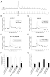
Inhibitors of CaMKs and PKA impair PTP at C1-B2 synapses. (A) Sample electrophysiological recording of PTP induction and decay at C1-B2 synapses in culture. Single action potentials in the C1 neuron are elicited at 0.05 Hz (lower trace) and the corresponding EPSPs evoked in the B2 neuron are simultaneously recorded (upper trace). After five basal stimuli, a tetanus (10 Hz for 2 seconds) is induced in the C1 neuron (arrowhead); 30 seconds later, the basal 0.05 Hz stimulation is resumed. The amplitude of the post-tetanic EPSPs is increased, reaching its peak 30 seconds after tetanus and progressively declining to pre-tetanic levels during the following 3-4 minutes. Bars, horizontal, 20 seconds; vertical, 8 mV upper trace, 60 mV lower trace. (B) Time-course of EPSP amplitude changes in two episodes of PTP evoked at a 30-minute interval in the same synapses. Values are normalized to the average amplitude of the last five pre-tetanic EPSPs. The peak amplitude and decay kinetics of PTP are nearly the same at t=0 (○) and at t=30 min (■) after tetanus. (C) Time-course of EPSP amplitude changes in two episodes of PTP evoked in the same synapses immediately before (○) and 30 minutes after bath application of the CaMKs inhibitor KN-93 (5 μM, ■). KN-93 nearly abolished the expression of PTP. (D) Time-course of the EPSP amplitude changes in episodes of PTP evoked in two distinct groups of synapses. In one group, the presynaptic C1 neuron was injected with the MLCK peptide (50-100 μM), an inhibitor of CaMKs (■). In the other group of synapses, the inactive MLCK control peptide was injected as a control (○). In both groups, PTP was evoked 30 minutes after injection of peptides. The MLCK peptide induces a dramatic impairment of PTP, similar to KN-93. (E) Time-course of EPSP amplitude changes in two episodes of PTP evoked in the same synapses immediately before (○) and 30 minutes after bath application of the PKA inhibitor Rp-cAMPS (500 μM, ■). (F) Mean peak PTP amplitudes measured at 30 seconds after tetanus in the experimental groups shown in panels B-E. Values are normalized to the mean peak potentiation measured under control conditions in each group. The peak amplitude of PTP is significantly reduced in the presence of KN-93, MLCK peptide and Rp-cAMPS. (G) Mean pre-tetanic EPSP amplitude in the PTP episodes shown in panels B-E. The various inhibitors used did not significantly alter the basal EPSP amplitude with respect to control conditions.
Fig. 3
Fig. 3
Presynaptic overexpression of GFP-tagged wild-type or mutant helSyn at C1-B2 synapses. (A) Phase-contrast micrograph of a C1-B2 soma-to-soma synapse in culture. (B) Epifluorescence micrograph of the same cell pair shown in A, 24 hours after the intracellular injection of mRNA encoding for GFP in the C1 neuron. GFP is expressed at high levels in the C1 neuron cytoplasm. At the focal plane of the picture, the dark profile of the C1 nucleus is visible (arrowheads), as well as the initial part of small neurites projecting from C1 onto the B2 surface (arrow). (C-H) Confocal stacks encompassing the whole volume of different C1-B2 cocultures overexpressing GFP (C,D), GFP-tagged wild-type helSyn (E,F) or GFP-tagged helSyn phosphorylation mutant (G,H). (C) GFP is distributed quite uniformly in the C1 cytoplasm and in the neurites growing onto the B2 surface. (D) Detail of the area of contact between the same C1 and B2 neurons shown in C. The arrow indicates a C1 neurite growing onto the B2 surface. (E) helSyn-GFP is more concentrated in discrete spots of increased fluorescence, which are particularly evident in the area of contact between the two cell bodies (arrowheads) and in varicose structures along the neurites (arrows). (F) Detail of the area of contact between the same C1 and B2 neurons shown in E. The arrow and arrowhead indicate sites of helSyn-GFP accumulation in the C1-B2 contact area and in neurites, respectively. (G) helSynALA9-GFP is strongly concentrated in numerous clusters widely distributed in the C1 cell body (double arrowhead) as well as in the C1-B2 contact area (arrowheads) and along C1 neurites growing onto B2 (arrow). (H) Detail of the area of contact between the same C1 and B2 neurons shown in G. The arrow and arrowhead indicate sites of helSynALA9-GFP accumulation in the C1-B2 contact area and in neurites, respectively. Note the higher fluorescence intensity of these helSynALA9-GFP puncta, as compared with the background fluorescence, with respect to the helSyn-GFP puncta shown in E and F. (I) Electron micrograph of a soma-to-soma C1-B2 synapse section encompassing the two cell bodies. The inset shows at higher magnification the meshwork of cellular processes in the contact area between the two somata. (L) Electron micrograph of a C1 neurite (asterisk) growing onto the surface of the postsynaptic B2 soma and containing a cluster of synaptic vesicles (arrowheads) at a putative synaptic site. (M) Detail of the synaptic site shown in L. Bars, 50 μm (A,B); 20 μm (C,E,G); 20 μm (D,F,H); 50 μm (I); 0.5 μm (L); 0.5 μm (M).
Fig. 4
Fig. 4
Presynaptic overexpression of the non-phosphorylatable helSynALA9 mutant impairs PTP at C1-B2 synapses. (A) Sample electrophysiological recording of EPSPs recorded in B2 neurons before (pre) and 30 seconds after (post) a tetanus was applied to presynaptic C1 neurons that were either untreated (control) or overexpressing either GFP (GFP), GFP-tagged wild-type helSyn (helSyn-GFP) or the GFP-tagged helSyn phosphorylation mutant (helSynALA9-GFP). Bars: horizontal, 2 seconds; vertical, 5 mV control, 4 mV GFP, 5 mV helSyn, 2.5 mV helSynALA9. (B) Time-course of EPSP amplitude changes in PTP episodes evoked by presynaptic tetanization (arrowhead) in the various experimental groups. Values are normalized to the average amplitude of the last five pre-tetanic EPSPs. The peak amplitude and decay kinetics of PTP are nearly the same in the GFP and helSyn group with respect to controls. Conversely, helSynALA9 overexpression determines a conspicuous impairment of PTP. (C) Mean amplitudes of the peak PTP measured at 30 seconds after tetanus in the experimental groups shown in B. Values are normalized to the mean value of peak potentiation measured in the control group. helSynALA9 overexpression dramatically reduces PTP to below 30% of control levels. (D,E) Amplitudes (D) and rise times (E) of pre-tetanic EPSPs in the experimental groups shown in B and C. No significant differences were detectable as a consequence of presynaptic GFP, helSyn-GFP or helSynALA9 overexpression.
Fig. 5
Fig. 5
Presynaptic overexpression of either wild-type or mutant helSyn does not affect facilitation or augmentation at C1-B2 synapses. (A) Sample electrophysiological recording of frequency facilitation at C1-B2 synapses during a train of five action potentials induced at 2 Hz in the presynaptic C1 neuron (lower trace). Note the progressive increase in the amplitude of the summating EPSPs recorded in the B2 neuron (upper trace). Bars: horizontal, 1.5 seconds; vertical, 5 mV upper trace, 20 mV lower trace. (B) The graph shows the mean facilitation index (expressed as the percent ratio between the fifth and the first EPSP during 2 Hz trains) measured in the different experimental groups. No significant change with respect to control conditions is observed after either helSyn-GFP or helSynALA9-GFP overexpression. (C) Sample electrophysiological recording of augmentation induction and decay at C1-B2 synapses. Single action potentials in the C1 neuron are elicited by intracellular depolarizing stimuli delivered at a basal frequency of 0.2 Hz (lower trace) and the corresponding EPSPs evoked in the postsynaptic B2 neuron are simultaneously recorded (upper trace). After some basal stimuli, a train of five action potentials at 10 Hz is induced in the C1 neuron (arrowhead). Two seconds after the train, the basal 0.2 Hz stimulation is resumed. The amplitude of the EPSPs after the train is increased for about 10 seconds. Bars: horizontal, 5 seconds; vertical, 5 mV upper trace, 20 mV lower trace. (D) Time-course of EPSP amplitude changes in augmentation episodes induced in synapses overexpressing either wild-type or mutant helSyn and in control synapses. No significant change in augmentation is observed after either helSyn-GFP or helSynALA9-GFP overexpression with respect to control conditions.

Similar articles

Cited by

References

    1. Abbott LF, Regehr WG. Synaptic computation. Nature. 2004;431:796–803. - PubMed
    1. Alle H, Jonas P, Geiger JR. PTP and LTP at a hippocampal mossy fiber interneuron synapse. Proc. Natl. Acad. Sci. USA. 2001;98:14708–14713. - PMC - PubMed
    1. Angers A, Fioravante D, Chin J, Cleary LJ, Bean AJ, Byrne JH. Serotonin stimulates phosphorylation of Aplysia synapsin and alters its subcellular distribution in sensory neurons. J. Neurosci. 2002;22:5412–5422. - PMC - PubMed
    1. Bonanomi D, Menegon A, Miccio A, Ferrari G, Corradi A, Kao HT, Benfenati F, Valtorta F. Phosphorylation of synapsin I by cAMP-dependent protein kinase controls synaptic vesicle dynamics in developing neurons. J. Neurosci. 2005;25:7299–7308. - PMC - PubMed
    1. Brager DH, Cai X, Thompson SM. Activity-dependent activation of presynaptic protein kinase C mediates post-tetanic potentiation. Nat. Neurosci. 2003;6:551–552. - PubMed

Publication types

MeSH terms

Substances