. 2007 Sep 4;104(36):14366-71.
doi: 10.1073/pnas.0703669104. Epub 2007 Aug 28.

Lsh controls Hox gene silencing during development

Affiliations

Lsh controls Hox gene silencing during development

Sichuan Xi et al. Proc Natl Acad Sci U S A. .

Erratum in

  • Proc Natl Acad Sci U S A. 2007 Oct 9;104(41):16389

Abstract

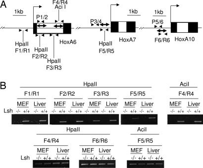
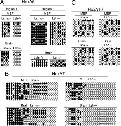
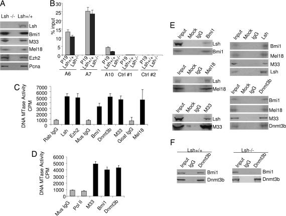
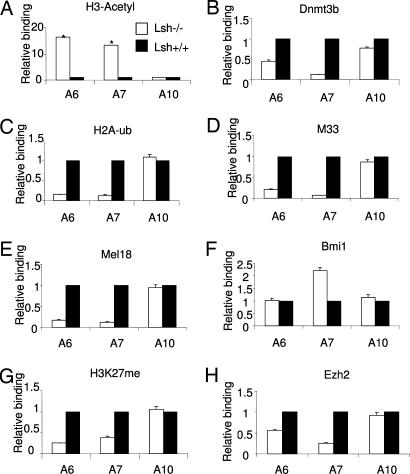
Polycomb-mediated repression and DNA methylation are important epigenetic mechanisms of gene silencing. Recent evidence suggests a functional link between the polycomb repressive complex (PRC) and Dnmts in cancer cells. Here we provide evidence that Lsh, a regulator of DNA methylation, is also involved in normal control of PRC-mediated silencing during embryogenesis. We demonstrate that Lsh, a SNF2 homolog, can associate with some Hox genes and regulates Dnmt3b binding, DNA methylation, and silencing of Hox genes during development. Moreover, Lsh can associate with PRC1 components and influence PRC-mediated histone modifications. Thus Lsh is part of a physiological feedback loop that reinforces DNA methylation and silencing of PRC targets.


Conflict of interest statement

The authors declare no conflict of interest.

Figures

Fig. 1.
Fig. 1.
Lsh deletion causes de-repression of Hox genes in various tissues. (A) RT-PCR analysis for detection of the indicated HoxA genes derived from Lsh−/− and Lsh+/+ MEFs, liver, brain, or whole embryo tissue (day 18 of gestation). (B) Real-time PCR analysis of HoxA gene expression comparing wild-type MEFs with Lsh−/− MEFs.
Fig. 2.
Fig. 2.
Decreased CpG methylation after Lsh deletion at selected Hox gene sites. (A) Map of the HoxA6, HoxA7, and HoxA10 genes illustrating the location of primers (black triangles). The methylation-sensitive restriction enzyme sites HpaII and AciI are shown, as well as the primers (designated F1/R1 to F6/R6) used for methylation-sensitive PCR analysis. The ChIP primers are designated P1/2, P3/4, and P5/6. The regions analyzed for bisulphate sequencing are indicated with a double arrow. (B) Methylation-sensitive PCR analysis using genomic DNA derived from Lsh−/− and Lsh+/+ MEFs or liver after digestion with HpaII or AciI. PCR analysis amplifying a region around an AciI site after HpaII digestion (or HpaII site after AciI digestion) served as a control for equal input of DNA. The primers F6/R6 amplify a region lacking methylation sensitive restriction enzyme sites.
Fig. 3.
Fig. 3.
Lsh deletion reduces DNA methylation at HoxA6 and HoxA7 sites. (A) Genomic DNA derived from Lsh−/− and Lsh+/+ MEFs or brain was subjected to bisulfite sequencing and examined at regions for two regions of the HoxA6 gene, as indicated in the map of Fig. 2A. Methylated CpG are presented by black circles and unmethylated sites by open circles. (B) Bisulfite sequencing analysis for MEFs and brain at the HoxA7 gene. (C) Bisulfite sequencing analysis for MEFs and brain at the HoxA10 gene.
Fig. 4.
Fig. 4.
Lsh is associated with PRC1 components. (A) Western blot analysis for detection of Bmi1, M33, Mel18, and Ezh2 using nuclear extracts derived from Lsh−/− and Lsh+/+ MEFs. Detection of Pcna and Lsh served as controls. (B) ChIP assays were performed on chromatin extracts derived from P19 (gray bar), Lsh+/+ (black bar), and Lsh−/− (open bar) MEFs using anti-Lsh antibodies and control IgG to detect association to specific HoxA6, HoxA7, and HoxA10 sites. Primers used for real-time PCR analysis are illustrated in Fig. 2A. In addition, two control primers were designed that are located within the HoxA cluster but >3,000 bp away from the HoxA10 or HoxA11 genes. The percentage of input was calculated for each precipitate. The values for the IgG control were <0.1% of input. (C and D) Nuclear extracts of P19 cells were immunoprecipitated with the indicated specific antibodies and assayed for DNA methyltransferase activity. (E) Western blot analysis for detection of Lsh after immunoprecipitation with anti-Bmi1, anti-Mel18, or anti-M33 antibodies using P19 nuclear extracts. Species-matched IgG or omission of antibody (Mock) served as controls. Western blot analysis for detection of Mel18, Bmi1, M33 after IP using anti-Lsh, or anti-Dnmt3b antibodies. (F) Western blot analysis for detection of Bmi1 after immunoprecipitation with anti-Dnmt3b comparing extracts derived from Lsh+/+ or Lsh−/− MEFs.
Fig. 5.
Fig. 5.
Lsh controls Dnmt3b recruitment and PRC-mediated histone modifications at Hox sites. ChIP assays were performed from chromatin extracts of Lsh−/− (open bar) and Lsh+/+ (black bar) MEFs using the indicated antibodies to detect specific histone modifications or association of specific proteins at HoxA genes. Primers used for real-time PCR analysis are illustrated in Fig. 2A. The percentage of input was calculated for each precipitate and the values expressed as ratio of Lsh−/− samples over wild type. The following antibodies were used: (A) Antiacetylation of H3. The asterix indicate the minimum ratio above wild-type controls, because the actual values for Lsh−/− samples exceeded the range of the standard curve. (B) Anti-Dnmt3b. (C) Anti-H2A-K116 ubiquitylation. (D) Anti-M33. (E) Anti-Mel18. (F) Anti-Bmi1. (G) Anti-H3-K27 trimethylation. (H) Anti-Ezh2.

References

    1. Jones PA. Semin Hematol. 2005;42:S3–8. - PubMed
    1. Goll MG, Bestor TH. Annu Rev Biochem. 2005;74:481–514. - PubMed
    1. Sparmann A, van Lohuizen M. Nat Rev Cancer. 2006;6:846–856. - PubMed
    1. Widschwendter M, Fiegl H, Egle D, Mueller-Holzner E, Spizzo G, Marth C, Weisenberger DJ, Campan M, Young J, Jacobs I, Laird PW. Nat Genet. 2007;39:157–158. - PubMed
    1. Schlesinger Y, Straussman R, Keshet I, Farkash S, Hecht M, Zimmerman J, Eden E, Yakhini Z, Ben-Shushan E, Reubinoff BE, et al. Nat Genet. 2007;39:232–236. - PubMed

Publication types