. 2007 Sep 4;104(36):14436-41.
doi: 10.1073/pnas.0702811104. Epub 2007 Aug 28.

CD200 and its receptor, CD200R, modulate bone mass via the differentiation of osteoclasts

Affiliations

CD200 and its receptor, CD200R, modulate bone mass via the differentiation of osteoclasts

Weiguo Cui et al. Proc Natl Acad Sci U S A. .

Abstract

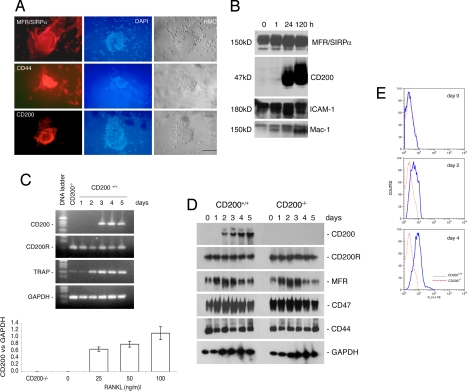
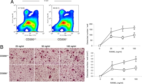
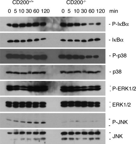
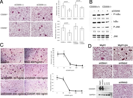
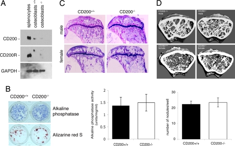
Fusion of macrophages is an essential step in the differentiation of osteoclasts, which play a central role in the development and remodeling of bone. Osteoclasts are important mediators of bone loss, which leads, for example, to osteoporosis. Macrophage fusion receptor/signal regulatory protein alpha (MFR/SIRPalpha) and its ligand CD47, which are members of the Ig superfamily (IgSF), have been implicated in the fusion of macrophages. We show that CD200, which is not expressed in cells that belong to the myeloid lineage, is strongly expressed in macrophages at the onset of fusion. By contrast, the CD200 receptor (CD200R), which, like CD200, belongs to the IgSF, is expressed only in cells that belong to the myeloid lineage, including osteoclasts, and in CD4+ T cells. Osteoclasts from CD200-/- mice differentiated at a reduced rate. Activation of the NF-kappaB and MAP kinase signaling pathways downstream of RANK, a receptor that plays a central role in the differentiation of osteoclasts, was depressed in these cells. A soluble recombinant protein that included the extracellular domain of CD200 rescued the fusion of CD200-/- macrophages and their activation downstream of RANK. Conversely, addition of a soluble recombinant protein that included the extracellular domain of CD200R or short-hairpin RNA-mediated silencing of the expression of CD200R prevented fusion. Thus CD200 engagement of the CD200R at the initiation of macrophage fusion regulated further differentiation to osteoclasts. Consistent with in vitro observations, CD200-/- mice contained fewer osteoclasts and accumulated more bone than CD200+/+ mice. The CD200-CD200R axis is therefore a putative regulator of bone mass, via the formation of osteoclasts.


Conflict of interest statement

The authors declare no conflict of interest.

Figures

Fig. 1.
Fig. 1.
Rat alveolar macrophages and mouse bone marrow-derived macrophages express CD200 upon multinucleation. (A) Freshly isolated rat alveolar macrophages were plated at confluency over 50% of the surface of each well to promote fusion and multinucleation. After 5 days, they were subjected to immunohistochemical analysis. Note that mononucleated macrophages were positive for MFR/SIRPα and CD44 but not for CD200. (Scale bar, 1 mm.) Also note that multinucleate rat alveolar macrophages contained hundreds of nuclei that were stained with DAPI (blue). (B) Freshly isolated rat alveolar macrophages were plated as in A and subjected to Western blot analysis at the indicated times. Note that CD200 was not detected in macrophages for the first 24 h. (C) Mouse bone marrow-derived macrophages were cultured in the presence of M-CSF (30 ng/ml) and RANKL (50 ng/ml) for the indicated times to induce the differentiation of multinucleate osteoclasts. Cells were analyzed by RT-PCR. Note that mouse bone marrow-derived macrophages expressed transcripts for CD200 receptor (CD200R) but not for CD200. The abundance of CD200 mRNA relative to that of GAPDH, in response to M-CSF (30 ng/ml) and increasing doses of RANKL, was determined. (Scale bars represent standard deviations; n = 3.) (D) Mouse bone marrow-derived macrophages were cultured in the presence of M-CSF (30 ng/ml) and RANKL (50 ng/ml) for the indicated times to induce the differentiation of multinucleate osteoclasts. Cells were subjected to Western blot analysis by using antibodies directed against the indicated antigens. (E) Flow-cytometric analysis (in a FACS) of the expression of CD200. Mouse bone marrow-derived macrophages were isolated from CD200+/+ and CD200−/− mice, cultured in the presence of M-CSF (30 ng/ml) and RANKL (50 ng/ml) and subjected to flow-cytometric analysis at the indicated times with an antibody directed against CD200 and a control isotype antibody. Bone marrow-derived macrophages expressed increasing amounts of CD200 with time in the presence of M-CSF and RANKL, which promote fusion, multinucleation and osteoclastogenesis.
Fig. 2.
Fig. 2.
Osteoclasts and their precursors are affected by the absence of CD200. (A) Bone marrow cells from 6-week-old CD200-deficient and wild-type mice were subjected to flow-cytometric analysis with antibodies directed against c-fms, Mac-1, and C-kit, as surface markers. Note that the absence of CD200 did not affect the number of osteoclast precursor cells (Left). (Scale bars, SD; n = 5.) (B) Bone-marrow-derived macrophages from 6-week-old CD200-deficient mice were cultured in the presence of M-CSF (30 ng/ml) and increasing concentrations of RANKL for 5 days to induce the differentiation of osteoclasts (Left). Bone marrow macrophages that lacked CD200 formed fewer osteoclasts than wild-type cells (Right). (Scale bars, SD; n = 5.)
Fig. 3.
Fig. 3.
In osteoclasts deficient in CD200, the activation of signaling molecules downstream of RANK is suppressed. Bone marrow macrophages isolated from CD200-deficient, and wild-type mice were cultured in the presence of M-CSF (5 ng/ml) for 12–18 h. Nonadherent cells were further cultured for 2 days in 24-well dishes, starved for 2 h, and then stimulated with 50 ng/ml RANKL for the indicated times. Cells were subjected to Western blot analysis with antibodies directed against the indicated antigens. The activation, by phosphorylation, of IκB and JNK was less extensive in cells that lacked CD200 than in wild-type cells. This experiment was repeated three times with similar results.
Fig. 4.
Fig. 4.
The CD200-CD200R axis is required for osteoclast fusion/multinucleation. (A) Bone-marrow-derived macrophages from 6-week-old wild-type mice were cultured in the presence of M-CSF (30 ng/ml) and RANKL (50 ng/ml) with or without the recombinant extracellular domain of CD200 (sCD200; 0.5 μg/ml) or recombinant prostate-specific antigen as a control. sCD200 allowed the differentiation of osteoclasts in macrophages that lacked CD200 (SD; n = 3). (B) Bone marrow macrophages isolated from CD200-deficient and wild-type mice were cultured in the presence of M-CSF (5 ng/ml) for 12–18 h. Nonadherent cells were cultured for a further 2 days in the presence of M-CSF (30 ng/ml), starved for 2 h, and then treated with RANKL (50 ng/ml) with or without sCD200 (0.5 μg/ml) for 30 min. The cells were then subjected to Western blot analysis with the indicated antibodies against IkBα and JNK and their phosphorylated forms. Numbers represent relative expression of P-IκB and P-JNK over JNK. The addition of sCD200 restored the activation of JNK but not of IκBα. (C) Bone-marrow-derived macrophages from 6-week-old wild-type mice were cultured in the presence of M-CSF (30 ng/ml) and RANKL (50 ng/ml) with or without the recombinant extracellular domain of the CD200 receptor (sCD200R). sCD200R blocked the fusion of macrophages (SD; n = 5), whereas recombinant prostate-specific antigen had no effect (osteoclast surface, 0.38 ± 0.1; osteoclast number, 170.0 ± 23.3). (D) Bone-marrow-derived macrophages from 6-week-old wild-type mice were cultured in the presence of M-CSF (30 ng/ml) for 2 days before being transduced with the retroviral vector MigR1, which encoded, or not, shRNAs designed after the CD200R1 cDNA. A construct encoding random (rdm) oligonucleotides was used as a control. Each of the three targeting retroviral constructs, namely shRNAi1, shRNAi2, and shRNAi3, abolished the expression of CD200R1 (see Western blot, Bottom Left) and prevented the formation of multinucleate osteoclasts (Top and Right). These experiments were reproduced several times with similar results.
Fig. 5.
Fig. 5.
The absence of CD200 increases bone density. (A) Bone marrow cells from 6- to 8-week-old CD200-deficient and wild-type mice were plated in 24-well plates (5 × 10⁶ cells/well) and cultured for 9–11 days in α-MEM supplemented with ascorbic acid (50 μg/ml) and β-glycerophosphate (10 mM) to acquire the osteoblast phenotype. Osteoblast lysates were analyzed for protein concentration and subjected to Western blot analysis with antibodies directed against mouse CD200, CD200R, and GAPDH. (B) Bone-marrow-derived osteoblasts were examined for ALP activity and stained for calcium with alizarin red S to allow quantitation of the number of nodules per well (SD; n = 6). Cell lysates were analyzed for ALP activity (Left; SD; n = 6). These experiments were repeated three times with similar results. (C) Toluidine blue-stained sections of proximal tibiae from 2-month-old CD200-deficient male and female mice and wild-type mice. (Scale bar, 1 mm.) (D) Microcomputed tomography analysis of distal femurs from 6-month-old male and female CD200-deficient mice. Note the increased density of trabeculae inside the distal femur of CD200-deficient male and female mice as compared with wild types. The widest diameter of the bone sections correspond ≈3 mm.

Similar articles

Cited by

References

    1. Vignery A. Int J Exp Pathol. 2000;81:291–304. - PMC - PubMed
    1. Vignery A. Trends Cell Biol. 2005;15:188–193. - PubMed
    1. Vignery A. J Exp Med. 2005;202:337–340. - PMC - PubMed
    1. Chen EH, Grote E, Mohler W, Vignery A. FEBS Lett. 2007;581:2181–2193. - PubMed
    1. Hernandez LD, Hoffman LR, Wolfsberg TG, White JM. Annu Rev Cell Dev Biol. 1996;12:627–661. - PubMed

Publication types