. 2007 Sep 4;104(36):14306-11.
doi: 10.1073/pnas.0701812104. Epub 2007 Aug 28.

Galphai generates multiple Pins activation states to link cortical polarity and spindle orientation in Drosophila neuroblasts

Affiliations

Galphai generates multiple Pins activation states to link cortical polarity and spindle orientation in Drosophila neuroblasts

Rick W Nipper et al. Proc Natl Acad Sci U S A. .

Abstract

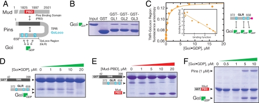
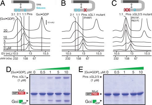
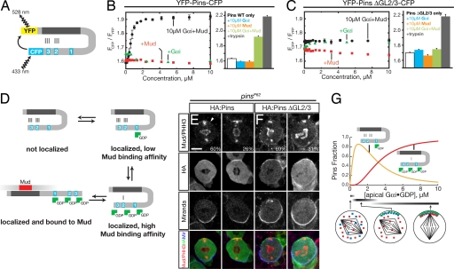
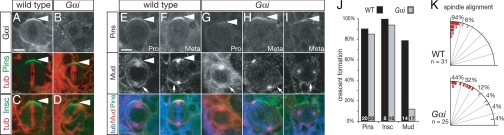
Drosophila neuroblasts divide asymmetrically by aligning their mitotic spindle with cortical cell polarity to generate distinct sibling cell types. Neuroblasts asymmetrically localize Galphai, Pins, and Mud proteins; Pins/Galphai direct cortical polarity, whereas Mud is required for spindle orientation. It is currently unknown how Galphai-Pins-Mud binding is regulated to link cortical polarity with spindle orientation. Here, we show that Pins forms a "closed" state via intramolecular GoLoco-tetratricopeptide repeat (TPR) interactions, which regulate Mud binding. Biochemical, genetic, and live imaging experiments show that Galphai binds to the first of three Pins GoLoco motifs to recruit Pins to the apical cortex without "opening" Pins or recruiting Mud. However, Galphai and Mud bind cooperatively to the Pins GoLocos 2/3 and tetratricopeptide repeat domains, respectively, thereby restricting Pins-Mud interaction to the apical cortex and fixing spindle orientation. We conclude that Pins has multiple activity states that generate cortical polarity and link it with mitotic spindle orientation.


Conflict of interest statement

The authors declare no conflict of interest.

Figures

Fig. 1.
Fig. 1.
Pins intramolecular interaction regulates Gαi and Mud binding. (A) Domain structure of Pins and intra- and intermolecular interactions (PBD, region of Mud that binds Pins). (B) Each of the Pins GoLoco motifs can bind Gαi. Individual GST fusions of the three GoLocos bind Gαi·GDP at qualitatively similar levels. (C) The Pins GoLocos are intrinsically independent, equivalent Gαi binding sites. The extent of Gαi·GDP binding to the Pins GoLocos was monitored by the fluorescence anisotropy of a tetramethylrhodamine attached to a cysteine at its C terminus. The curve represents a model with three equivalent, independent binding sites of affinity Kd = 530 ± 80 nM. A Scatchard analysis is shown in Inset where the binding function is equal to the concentration of Pins-bound Gαi·GDP divided by the total concentration of Pins. (D) Gαi disrupts the Pins intramolecular interaction. In a qualitative “pull-down” assay, Gαi·GDP competes with the Pins GLR for binding to the Pins TPRs. Although Gαi ultimately disrupts the TPR–GLR interaction, at intermediate Gαi concentrations (5–10 μM), a Gαi–GLR–TPR complex can be formed, presumably those that result in occupation of GoLoco1 but not GoLoco2/3. At a higher concentration (20 μM), occupation of all three GoLoco motifs by Gαi interferes with the interaction of the GLR with the TPR region. Proteins are stained with Coomassie brilliant blue. (E) The Mud PBD disrupts the Pins intramolecular interaction. Binding of Mud to the Pins TPRs (as in B) competes with the Pins GoLocos. (F) Gαi increases the affinity of Pins for Mud. Full-length Pins binds weakly to the Mud PBD, but binding is enhanced by the presence of Gαi·GDP, indicating that Gαi and Mud bind cooperatively to Pins.
Fig. 2.
Fig. 2.
Differential repression of the three GoLocos by the Pins intramolecular interaction. (A) Analysis of Pins binding to Gαi·GDP by gel-filtration chromatography. Pins and mixtures of Pins and Gαi·GDP were separated by gel filtration. Marks indicating the elution volumes of 2:1 and 1:1 Gαi:Pins complexes were determined by using single and double Pins GoLoco mutants, respectively. The column elution volumes (EV) of standard proteins to give a calibrated molecular weight (cMW) are shown on the x axis. The protein composition of the 20 μM Gαi eluate for this and B and C are shown in SI Fig. 5C. (B) Analysis of Pins with an inactive GoLoco1 (Pins ΔGL1) binding to Gαi·GDP by gel-filtration chromatography. Loss of GoLoco1 causes loss of the high-affinity peak that occurs at low Gαi concentrations. (C) Analysis of Pins with an inactive GoLoco 2 and 3 (Pins ΔGL2/3) binding to Gαi·GDP by gel-filtration chromatography. Only the high-affinity interaction remains after loss of GoLocos 2 and 3. (D) Cooperative binding of Gαi and Mud to Pins does not require GoLoco1. In the absence of GoLoco1, Gαi·GDP enhances the affinity of Pins for Mud. (E) GoLocos 2 and 3 are required for cooperative binding of Gαi and Mud to Pins. Although Gαi·GDP can bind to Pins in which GoLocos 2 and 3 are inactivated (C), binding does not lead to cooperative Mud binding.
Fig. 3.
Fig. 3.
Pins undergoes a conformational change into a high Mud binding affinity state in response to Gαi binding to GoLocos 2 and 3. (A) Architecture of the Pins FRET sensor. (B) Full transition to the Pins open state requires both Gαi·GDP and Mud. Shown in the bar graph are the FRET ratios for the WT Pins FRET sensor ([YFP–Pins–CFP] = 200 nM) with various combinations of Gαi·GDP and Mud ligands present at 10 μM concentration and after trypsin digestion. Error bars equal one SD (n = 3). (C) Pins with inactivated GoLocos 2 and 3 is unable to transition to open state with Gαi·GDP and Mud present. Shown in the bar graph are the FRET ratios for the Pins ΔGL2/3 FRET sensor ([YFP–Pins–CFP] = 200 nM) with various combinations of Gαi·GDP and Mud ligands present at 10 μM concentration. Error bars equal one SD (n = 3). (D) Model for coupled Gαi·GDP and Mud binding and relationship to Pins conformational states. (E) HA:Pins can rescue Mud localization in pins larval neuroblasts. Pins localization is detected by anti-HA antibody, Mud is detected by anti-Mud antibody, and the mitotic stage is determined by anti-phospho histone H3 (α-PHH3) antibody. Moderate (60%) or strong (26%) Mud apical crescents were observed in metaphase neuroblasts. The localization of Mud to the spindle poles is independent of Pins activity (13). Miranda was used as a basal marker. (F) Pins with inactivated GoLocos 2 and 3 fails to rescue Mud apical enrichment in pins larval neuroblasts. Staining is as in E. Although HA:Pins ΔGL2/3 is correctly localized, only weak or moderate Mud apical staining was observed in 31% of metaphase neuroblasts. (Scale bar: E and F, 5 μm.) (G) Pins–Gαi response profile. The concentration of Pins with Gαi bound at GoLoco1 and Gαi-saturated Pins is shown as a function of Gαi concentration based on the model shown in D. This model uses the intrinsic affinity of Gαi for the GoLocos (Kd = 530 nM) and assumes that the inactive form of Pins is favored 1,000:1 over active Pins in the absence of Gαi or Mud. Simulations were performed with Berkeley Madonna. Predicted neuroblast phenotypes with the state of cortical polarity (red, Mud; blue, Pins; green, Gαi) and spindle positioning are shown at low, medium, and high Gαi concentration ranges.
Fig. 4.
Fig. 4.
Neuroblasts with reduced Gαi levels can recruit Pins/Insc but not Mud to the apical cortex and have spindle alignment defects. (A and B Top) Gαi apical cortical protein levels are reduced in Gαi zygotic mutant larval neuroblasts (B) compared with WT (A). (A and B Lower) Same neuroblast labeled for α-tubulin (tub) and Pins. Strong Pins crescents were observed in both WT (Left) and zygotic Gαi mutant (Right) metaphase neuroblasts. (C and D) Strong Insc crescents were observed in both WT (C) and zygotic Gαi mutant (D) metaphase neuroblasts (cortical fluorescence intensities Insc 84% of WT; see Methods for details). (E–I) Gαi zygotic mutant larval neuroblasts have robust Pins apical crescents but a loss of Mud apical protein crescents (cortical fluorescence intensities of Pins 90% of WT; see Methods for details). (E and F) WT larval neuroblasts have apical crescents of Pins and Mud at prophase (pro) and metaphase (meta) (arrowheads), as well as strong Mud staining on centrosomes and at the basal cortex (arrows). (G–I) Gαi mutant larval neuroblasts show Pins apical crescents but have defects in forming Mud apical protein crescents at prophase and metaphase (arrowheads), although Mud at the basal cortex is unaffected (arrows). (Scale bar: E–I, 5 μm.) (J) Quantification of apical crescent formation at metaphase in WT and Gαi mutant neuroblasts (see Methods for details). The number in each bar represents the number of neuroblasts examined. (K) Gαi zygotic mutant neuroblasts show spindle alignment defects. Quantification of apical spindle pole alignment (red ticks) relative to the center of the Pins cortical crescent (vertical line). WT spindles are tightly aligned, but Gαi mutant spindles are frequently misaligned.

Similar articles

Cited by

References

    1. Betschinger J, Knoblich JA. Curr Biol. 2004;14:R674–R685. - PubMed
    1. Cowan CR, Hyman AA. Annu Rev Cell Dev Biol. 2004;20:427–453. - PubMed
    1. Yu F, Kuo CT, Jan YN. Neuron. 2006;51:13–20. - PubMed
    1. Wodarz A. Curr Opin Cell Biol. 2005;17:475–481. - PubMed
    1. Morrison SJ, Kimble J. Nature. 2006;441:1068–1074. - PubMed

Publication types

MeSH terms

Substances