. 2007 Sep 4;104(36):14489-94.
doi: 10.1073/pnas.0701311104. Epub 2007 Aug 28.

Essential role for autophagy protein Atg7 in the maintenance of axonal homeostasis and the prevention of axonal degeneration

Affiliations

Essential role for autophagy protein Atg7 in the maintenance of axonal homeostasis and the prevention of axonal degeneration

Masaaki Komatsu et al. Proc Natl Acad Sci U S A. .

Abstract

Autophagy is a regulated lysosomal degradation process that involves autophagosome formation and transport. Although recent evidence indicates that basal levels of autophagy protect against neurodegeneration, the exact mechanism whereby this occurs is not known. By using conditional knockout mutant mice, we report that neuronal autophagy is particularly important for the maintenance of local homeostasis of axon terminals and protection against axonal degeneration. We show that specific ablation of an essential autophagy gene, Atg7, in Purkinje cells initially causes cell-autonomous, progressive dystrophy (manifested by axonal swellings) and degeneration of the axon terminals. Consistent with suppression of autophagy, no autophagosomes are observed in these dystrophic swellings, which is in contrast to accumulation of autophagosomes in the axonal dystrophic swellings under pathological conditions. Axonal dystrophy of mutant Purkinje cells proceeds with little sign of dendritic or spine atrophy, indicating that axon terminals are much more vulnerable to autophagy impairment than dendrites. This early pathological event in the axons is followed by cell-autonomous Purkinje cell death and mouse behavioral deficits. Furthermore, ultrastructural analyses of mutant Purkinje cells reveal an accumulation of aberrant membrane structures in the axonal dystrophic swellings. Finally, we observe double-membrane vacuole-like structures in wild-type Purkinje cell axons, whereas these structures are abolished in mutant Purkinje cell axons. Thus, we conclude that the autophagy protein Atg7 is required for membrane trafficking and turnover in the axons. Our study implicates impairment of axonal autophagy as a possible mechanism for axonopathy associated with neurodegeneration.


Conflict of interest statement

The authors declare no conflict of interest.

Figures
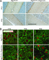
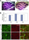
Fig. 1.
Fig. 1.
Deletion of Atg7 specifically in Purkinje cells caused progressive dystrophic swelling of axon terminals. (A) Immunohistochemistry of Atg7 protein expression in Purkinje cells of Atg7flox/flox and Atg7flox/flox;Pcp2-Cre mice at P15 and P19. The endogenous Atg7 protein was present at P15 but absent at P19 in the Atg7flox/flox;Pcp2-Cre Purkinje cells. (Scale bar: 100 μm.) (B) Progression of the abnormal Purkinje cell axon terminal swellings in the DCN of Atg7flox/flox;Pcp2-Cre mice (anti-calbindin immunofluorescent staining in green with anti-NeuN counterstained in red) at P19, P35, and P56. Atg7flox/flox was used as control. (Scale bar: 20 μm.) n = 3–5.
Fig. 2.
Fig. 2.
The axonal dystrophic swellings of the Atg7-deficient Purkinje cells contained no GFP-LC3 labeled autophagosomes but accumulated p62/SQSTM1. (A) The absence of GFP-LC3 puncta in Purkinje cell axonal dystrophic swellings (b and c) and somata (f and g) of Atg7flox/flox;Pcp2-Cre/GFP-LC3 mice (P35). GFP-LC3 puncta were found in GFP-LC3/Lurcher Purkinje cell axonal dystrophic swellings (d) and somata (h) (P12). DCN (a) and Purkinje cell layer (PCL) (e) of control mice Atg7flox/flox/GFP-LC3 are shown. (Scale bars: a, b, e, and f, 20 μm; c, d, g, and h, 10 μm.) (B) Anti-p62/SQSTM1 immunofluorescent staining (in green) showed accumulation of p62/SQSTM1 in Purkinje cell axonal dystrophic swellings (calbindin labeling in red) (white arrows) in the DCN of Atg7flox/flox;Pcp2-Cre mice at P56. Atg7flox/flox was used as control. (Scale bar: 10 μm.)
Fig. 3.
Fig. 3.
Deletion of Atg7 in Purkinje cells had little effect on the morphology of cerebellar cortex, Purkinje cell dendritic tree and spines in Atg7flox/flox;Pcp2-Cre mice at P56. (A) H&E-stained images of midsagittal sections from Atg7flox/flox and Atg7flox/flox;Pcp2-Cre cerebella at P56. (Scale bar: 0.5 mm.) n = 3–5. (B) Quantification of the molecular layer thickness (as the distance between lobules V and VI of the Purkinje cell layer divided by 2) from the cerebellar midsagittal sections of Atg7flox/flox and Atg7flox/flox;Pcp2-Cre mice at P19, P35, and P56. n = 3–5. (C) Immunofluorescent staining of cerebellar midsagittal sections shows normal localization and appearance of mGluR1α (in red) in Atg7flox/flox;Pcp2-Cre mice compared with Atg7flox/flox mice at P56. Green indicates anti-calbindin. (Scale bar: 10 μm.) n = 3.
Fig. 4.
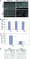
Fig. 4.
Time course of Purkinje cell degeneration and locomotive behavioral deficits in Atg7flox/flox;Pcp2-Cre mice. (A) Anti-calbindin immunofluorescent staining of the cerebellar midsagittal sections of Atg7flox/flox and Atg7flox/flox;Pcp2-Cre mice at P35 and P56. (B) Quantitation of Purkinje cells at lobules IV–V of the midsagittal sections of Atg7flox/flox and Atg7flox/flox;Pcp2-Cre mice at P19, P35, and P56 based on H&E-stained images. n = 3, 3, and 3 for Atg7flox/flox at P19, P35, and P56, respectively. n = 2, 3, and 5 for Atg7flox/flox;Pcp2-Cre at P19, P35, and P56, respectively (*, P < 0.0005). (C) At P56, Atg7flox/flox and Atg7flox/flox;Pcp2-Cre mice showed no significant difference in the time spent on the rod in rotarod assay. At 1 year, Atg7flox/flox mice spent much longer time on the rod than Atg7flox/flox;Pcp2-Cre mice (**, P < 0.05). (D) In gait analyses at P56, Atg7flox/flox and Atg7flox/flox;Pcp2-Cre mice showed similar step width and overlapping of forefeet and hindfeet (forefeet, red; hindfeet, black). At 1 year old, Atg7flox/flox;Pcp2-Cre mice showed shorter step width than Atg7flox/flox mice as well as nonoverlapping forefeet and hindfeet (left feet, black; right feet, red). For both C and D, n = 5 at P56; n = 3 and 4 at 1 year.
Fig. 5.
Fig. 5.
Deletion of Atg7 in Purkinje cells led to aberrant membrane structures in the axonal dystrophic swellings. Ultrastructural image of normal myelinated Purkinje cell axons (white arrows) and axon terminals (black arrows) in the DCN of Atg7flox/flox mice (A) and Purkinje cell axonal dystrophic swellings (black arrows) in the DCN of Atg7flox/flox;Pcp2-Cre mice (B–F). (B and E) Stacks of cisternal membranes (white arrows). (C and D) Convoluted double-membrane whorls (white arrows). (F) The arrays of abnormal filaments (white arrows). (G) A dystrophic axon (black arrows) of Lurcher Purkinje cells containing numerous autophagosomes. (Scale bars: 500 nm.)
Fig. 6.
Fig. 6.
Deletion of Atg7 in Purkinje cells abolished double-membrane vacuole-like structures in their axon terminals in the DCN. (A) Ultrastructural images show the presence of vacuole-like structures with double membranes in Purkinje cell preterminal axons of Atg7flox/flox mice (a–e, white arrows) and the abolishment of these structures in Atg7flox/flox;Pcp2-Cre mice (f) at P35. (Scale bars: 0.5 μm.) (B) Comparison of the numbers of these vacuole-like structures per transmission electron microscopy micrograph (50 μm2) in the DCN of Atg7flox/flox;Pcp2-Cre versus Atg7flox/flox mice (ratio, 4.0; P = 0.00003). (C) Comparison of the volume fraction of double-membrane vacuole-like structures by point counting of transmission electron microscopy micrographs in the DCN of Atg7flox/flox;Pcp2-Cre versus Atg7flox/flox mice (ratio, 6.9; P = 0.00002).

Similar articles

Cited by

References

    1. Levine B, Klionsky DJ. Dev Cell. 2004;6:463–477. - PubMed
    1. Mizushima N, Yamamoto A, Matsui M, Yoshimori T, Ohsumi Y. Mol Biol Cell. 2004;15:1101–1111. - PMC - PubMed
    1. Rubinszstein DC, DiFiglia M, Heintz N, Nixon RA, Qin ZH, Ravikumar B, Stefanis L, Tolkovsky A. Autophagy. 2005;1:11–22. - PubMed
    1. Yue Z, Horton A, Bravin M, DeJager PL, Selimi F, Heintz N. Neuron. 2002;35:921–933. - PubMed
    1. Dixon JS. Nature. 1967;215:657–658. - PubMed

Publication types

MeSH terms