. 2007 Aug 29;2(8):e787.
doi: 10.1371/journal.pone.0000787.

Insights from amphioxus into the evolution of vertebrate cartilage

Affiliations

Insights from amphioxus into the evolution of vertebrate cartilage

Daniel Meulemans et al. PLoS One. .

Abstract

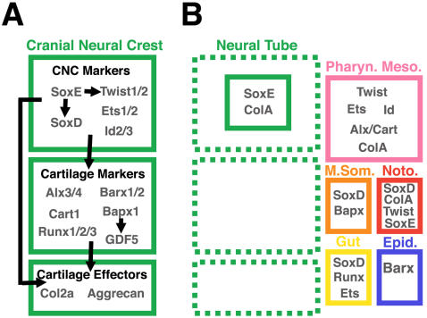
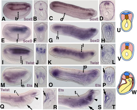
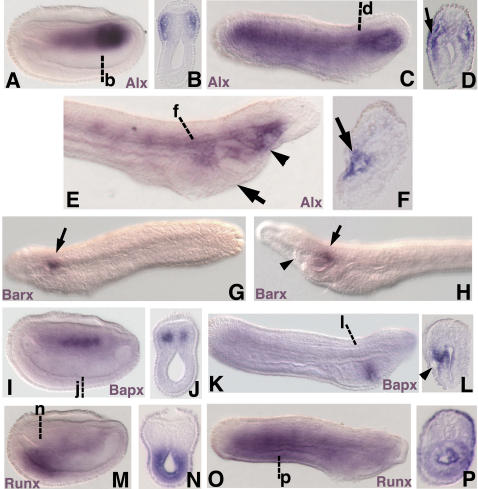
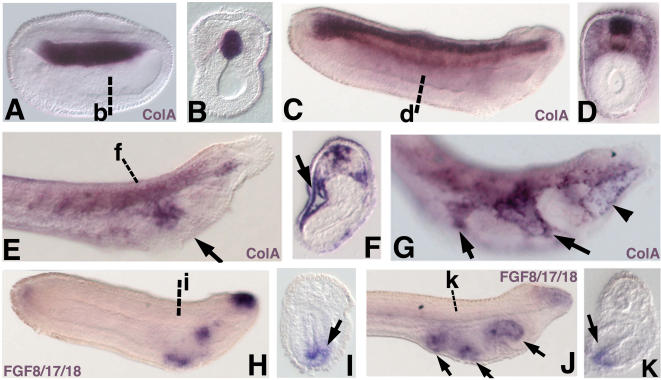
Central to the story of vertebrate evolution is the origin of the vertebrate head, a problem difficult to approach using paleontology and comparative morphology due to a lack of unambiguous intermediate forms. Embryologically, much of the vertebrate head is derived from two ectodermal tissues, the neural crest and cranial placodes. Recent work in protochordates suggests the first chordates possessed migratory neural tube cells with some features of neural crest cells. However, it is unclear how and when these cells acquired the ability to form cellular cartilage, a cell type unique to vertebrates. It has been variously proposed that the neural crest acquired chondrogenic ability by recruiting proto-chondrogenic gene programs deployed in the neural tube, pharynx, and notochord. To test these hypotheses we examined the expression of 11 amphioxus orthologs of genes involved in neural crest chondrogenesis. Consistent with cellular cartilage as a vertebrate novelty, we find that no single amphioxus tissue co-expresses all or most of these genes. However, most are variously co-expressed in mesodermal derivatives. Our results suggest that neural crest-derived cartilage evolved by serial cooption of genes which functioned primitively in mesoderm.


Conflict of interest statement

Competing Interests: The authors have declared that no competing interests exist.

Figures

Figure 1
Figure 1. A provisional gene network operating in nascent neural crest-derived cartilage and expression of network component homologs in amphioxus.
(A) We have classified genes in the network as cranial neural crest (CNC) markers, cartilage markers, or effector genes based on their expression, regulatory relationships, and biochemical functions. Among the CNC markers are Sox9 (SoxE), Sox5/6 (SoxD), Twist1/2 and Ets1/2 genes. All of these factors are expressed in post-migratory chondrogenic cranial neural crest , , , , . SoxE, SoxD, and Twist1/2 have been shown to cross-regulate, and to activate cartilage specifiers and effector genes. SoxE is required for expression of both SoxD and Twist1/2 in migrating CNC , , while Twist1/2 is necessary for the continued expression of SoxE in postmigratory CNC. Both SoxE and SoxD cooperate to directly activate the definitive cartilage differentiation marker Col2a1 in chondroblasts, while Twist1/2 is required for expression of the aristalless-related transcription factors Alx3/4 and Cart1 . Ets1/2 expression overlaps temporally and spatially with SoxE, SoxD and Twist1/2, though functional relationships between it and the other network components have yet to be demonstrated . In sea urchins, Ets1/2 and Alx3/4 orthologs are necessary for the formation of skeletogenic mesenchyme and are regulated by the same upstream factors, suggesting they cooperate in an evolutionarily ancient skeletogenic program , . As chondrogenesis begins, presumptive pharyngeal chondrocytes express genes grouped here as cartilage markers (Barx1/2, Alx3/4, Cart1, Runx1/2/3, Bapx1, and GDF5). These genes are expressed in differentiating CNC-derived chondrocytes –, , , –, are downstream of CNC specfiers and upstream of effector genes like Col2a1 and Aggrecan, Barx1 physically interacts with Sox9 to directly activate Collagen2a1 expression . As indicated above, Alx3/4 and Cart1 are regulated by Twist1/2 . Runx1/2/3 expression in chondrocytes is dependent on SoxE function. In the pharynx, Bapx1 functions mainly to position the jaw joint by regulating expression of GDF5 , . In the mesoderm-derived axial skeleton, however, Bapx1 is expressed broadly and operates upstream of Sox9, Col2a1, and Runx1/2/3 , . Essential for maintenance and establishment of the chondrogenic subnetwork are signaling molecules of the FGF and Endothelin families which are secreted by surround pharyngeal endoderm and ectoderm, (not shown). These genes also mediate pharyngeal arch patterning by activating nested expression of various transcription factors in the nascent cartilages including Dlx and Msx genes, Gsc, and Hand2 , (not shown). (B) The major expression domains of chondrogenic neural crest gene homologs in amphioxus neurulae and larvae. No single cell type expresses the complete set of vertebrate chondrogenic network genes, indicating the cranial neural crest cartilage program is a vertebrate novelty. Notably, most factors are expressed in mesodermal derivatives, suggesting neural crest-derived cartilage evolved via repeated cooption of primitively mesodermal genes.
Figure 2
Figure 2. Expression of amphioxus SoxE, SoxD, Twist, and Ets at late neurula (15 h) and larval stages.
In all panels showing wholemount specimens, anterior is to the left. (A) SoxE expression in ventral notochord and medial neural plate in late neurula. (B) Section through b in A. Superficial ectoderm staining is caused by adhesion of precipitate forming during the in situ hybiridzation procedure to the outside of the embryo. This artefact is distinguishable from actual signal because it is acellular, not visible in wholemount, and only readily apparent in overstained sections viewed using phase contrast optics. (C) SoxE expression throughout the neural tube and in ventral notochord cells at the anterior and posterior tips in early larva (24 h). (D) Section through d in C showing neural tube staining. (E) SoxD expression in the medial somites and notochord in late neurula. (F) Section through f in E. (G) SoxD expression in the posterior notochord, anterior gut, and cerebral vesicle of early larva. (H) Section through h in G showing notochord expression. (I)Twist expression in lateral somites and notochord in late neurula (J) Section through j in I. (K) Twist expression in ventrolateral mesoderm of early larva. (L) Section through the pharynx at l in K showing expression in pharyngeal mesoderm (arrow). (M) Ets expression in the posterior gut, anterior notochord, and ventral aspect of the anterior somites of late neurula. (N) Section through n in M. (O) Ets expression in the gut and anterior mesendoderm of early larva. (P) Section through the pharynx at p in O showing expression in pharyngeal mesoderm (arrow) and gut. (Q) Twist expression in the mesoderm of the first pharyngeal arch (arrow) and right gut diverticulum (arrowhead) of 1.5d larva. (R) Section through the first pharyngeal arch at r in Q showing mesodermal expression (arrow). (S) Ets expression in the mesoderm of the first pharyngeal arch (arrow), gut diverticulae (arrowhead), and cerebral vesicle of 1.5d larva. (T) Section through the first pharyngeal arch at t in S showing mesodermal expression (arrow). (U) Diagram of cross section midway through late neurula. (V) Diagram of cross section midway through early larva. (W) Diagram of cross section through first pharyngeal arch in 1.5d larva. In cephalochordate larvae, gill slits on opposite sides of the pharynx form asynchronously, with the right gill slits forming first. Thus, cross sections through the pharynx of amphioxus larvae reveal single gill bars rather than the symmetrical pharyngeal arches typical of analogous sections through vertebrate embryos. In U,V, and W, light blue is epidermal ectoderm, dark blue is the neural tube, brown is the notochord, yellow is endoderm, pink is somitic mesoderm, and red is pharyngeal arch mesoderm.
Figure 3
Figure 3. Expression of amphioxus Alx, Barx, Bapx, and Runx in late neurulae (15 h) and larvae.
In all panels showing wholemount specimens, anterior is to the left unless otherwise indicated. (A) Alx expression in the lateral somites and gut diverticulae of late neurula. Strongest expression is seen in the gut diverticulae and first somites. (B) Section through the first somites at b in A. (C) Alx expression in ventral mesoderm and the anterior gut diverticulae of early larva (24 h). (D) Section through the pharynx at d in C showing expression in the pharyngeal mesoderm (arrow). (E) Alx expression in the mesoderm of the first pharyngeal arch (arrow) and the right gut diverticulum (arrowhead) of 1.5d larva. (F) Section through the first pharyngeal arch at f in E showing expression in mesoderm. (G) Anterior is to the left. Left side of an early larva focused in the plane of the ectoderm showing Barx expression in a patch of ectoderm (arrow) just caudal to the forming preoral pit. (H) Anterior is to the left. Left side of a 1.5d larva focused in the plane of the ectoderm showing Barx expression in a few ectodermal cells (arrow) caudal to the preoral pit (arrowhead). (I) Bapx expression in the medial somites of late neurula. (J) Section through j in I. (K) Bapx expression in a stripe of pharyngeal endoderm on the right side of an early larva approximating the region of the nascent first gill slit. (L) Section through l in K showing endodermal staining (arrowhead). (M) Runx expression in the posterior gut of late neurula. (N) Section through n in M. (O) Runx expression in the gut of early larva. (P) Section through p in O.
Figure 4
Figure 4. Expression of amphioxus ColA, and FGF8/17/18 in late neurulae (15 h) and larvae.
In all panels showing wholemount specimens, anterior is to the left. (A) ColA expression in the nascent notochord in late neurula. (B) Section at the level of b in A. (C) ColA expression in the neural tube and notochord in early larva (24 h). (D) Section through d in C showing weak expression in somitic mesoderm. (E) ColA expression in the mesoderm of the first pharyngeal arch (arrow) of 1.5d larva. (F) Section through the first pharyngeal arch at f in E showing mesodermal expression (arrow). (G) ColA expression in 2 gill slit larva. Strong expression is seen in the mesoderm of the first and second pharyngeal arches (arrows) and in individual cells of the right gut diverticulum (arrowhead). (H) FGF8/17/18 expression in dorsal anterior ectoderm and two patches of pharyngeal endoderm. (I) Section through the pharynx at i in H showing FGF8/17/18 expression in ventral endoderm (arrow). (J) FGF8/17/18 expression in the pharyngeal endoderm of 1.5d larva (arrows). (K) Section through k in J showing expression in ventral endoderm (arrow).

Similar articles

Cited by

References

    1. Couly GF, Coltey PM, Le Douarin NM. The triple origin of skull in higher vertebrates: a study in quail-chick chimeras. Development. 1993;117:409–429. - PubMed
    1. Meulemans D, Bronner-Fraser M. Central role of gene cooption in neural crest evolution. J Exp Zoolog B Mol Dev Evol. 2005;304:298–303. - PubMed
    1. Jeffery WR, Strickler AG, Yamamoto Y. Migratory neural crest-like cells form body pigmentation in a urochordate embryo. Nature. 2004;431:696–699. - PubMed
    1. Gans C, Northcutt RG. Neural Crest and the Origin of Vertebrates-a New Head. Science. 1983;220:268–274. - PubMed
    1. Northcutt RG, Gans C. The genesis of neural crest and epidermal placodes: a reinterpretation of vertebrate origins. Q Rev Biol. 1983;58:1–28. - PubMed

Publication types